Keywords
Waardenberg syndrome, Autosomal dominant, Dystopia canthorum, Heterochromia, Poliosis, Hypertelorism
Introduction
Waardenberg syndrome is a rare genetic condition characterised by pigmentary and auditory disorder. Even though it was first reported by P.J Waardenburg, Van Der Hoeve is credited with first describing the condition. The first cases of patients with similar clinical phenotype showed markedly increased distance between inner angles of the eyelids (37-52 or more in adults). The interpapillary distance and outer canthal distance however are within normal limits, hence the nasal part of the ocular surface shows almost no sclera [1].
Pigmentary disorders occur due to failure of differentiation of melanocytes. It can present in many forms of clinical signs. In the first report case, the pigmentary disorder was heterochromia, but further study report other kind of pigmentary disorders , such as skin hypopigmentation and hair (polyosis). Shields, Carol et al (2013) investigated further ocular pigmentary disorders and found not only changes affecting the iris, but also affecting the choroid (hypopigmentation of the choroid) [2,3].
Sensoryneural deafness is one of the symptoms most likely to be present in patient with Waardenberg syndrome. It was described by P.J Waardenberg in the original case. In further studies, this symptom become a major criteria to differentiate the type of Waardenberg syndrome [1,2].
In 1995, Liu et al. describe the chromosomal abnormality that contribute to the clinical manifestations of Waardenberg syndrome. Based on this studies, waardenberg syndrome can be divided into 2 types. Type 1 and type 2, differentiated by the intercanthal distance and the broad nasal bridge [2,4,5].
Case Report
This study represented 10 family members, 7 among them are presence with major criteria of waardenberg syndrome, while 3 of them presence with the minor criteria. All the 10 members of the family are divided into 4 generations. There was no consanguineous marriage among the family members.
Case 1
60 years old woman with an abnormal pigmentation of the eye since birth. There is a history of blurry vision since more than 20 years durations but no history of using spectacles. There was history of white hair from a young age, but no presentation of hypopigmentation lessions on the skin. The patient was examined by ENT and concluded the patient was suffered from sensory neural deafness. There is also a history of hearing loss in the family (Figure 1).
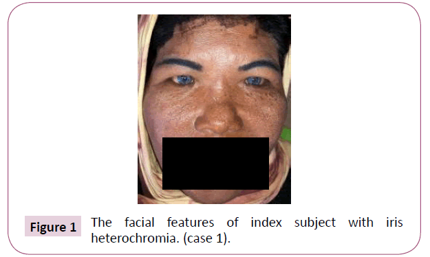
Figure 1: The facial features of index subject with iris heterochromia. (case 1).
The uncorrected visual acuity of the patient at presentation was 20/50 on both eyes with best corrected visual acuity 20/20 with+1.25 Spher on both eyes. The intraocular pressure on both eyes was 19mmHg.
The inner intercanthal distance is 42, with pupil distance 63/60. The wide intercathal distance with a normal range of pupil distance is the sign of hypertelorism which in this patient it might be occurs due to widening of nasal bridge. Her late father and late grandfather also have the same facial features.
Case 2
One daughter of the index subject also presence with bright irides, hypopigmentation of the skin and broad nasal bridge. She has been assessed with ENT and diagnosed with sensory neural deafness (Figure 2).
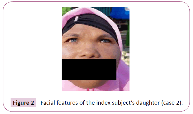
Figure 2: Facial features of the index subject’s daughter (case 2).
Case 3
The grandson of the index subject also presence with heterochromia iris. The sensorineural function of the grandson couldn’t be assessed during the study. There were no speech delayed presence. The baby was born from a mother whose also has the same facial features (Figure 3).
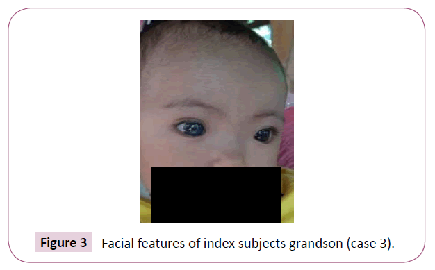
Figure 3: Facial features of index subjects grandson (case 3).
Case 4
Younger sibling of the index subject also presence with bright irides, hypopigmentation of the skin and broad nasal bridge. She has been assessed with ENT and diagnosed with sensory neural deafness (Figure 4).
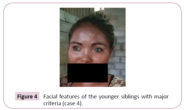
Figure 4: Facial features of the younger siblings with major criteria (case 4).
Case 5
The same condition can be found in the family. The index subject’s siblings also suffered from waardenberg syndrome. The younger siblings of the patient show poliosis and congenital sensorineural deafness which are the major criteria of waardenberg syndrome. Those conditions accompanied by broad and high nasal root and hypo pigmented on the skin which represents the minor criteria of waardenberg syndrome (Figure 5).
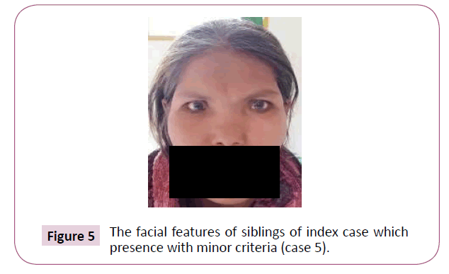
Figure 5: The facial features of siblings of index case which presence with minor criteria (case 5).
Case 6
The brother of siblings of the patient shows poliosis and congenital sensorineural deafness which are the major criteria of waardenberg syndrome. Those conditions accompanied by broad and high nasal root, synophris and hypo pigmented on the skin which represents the minor criteria of waardenberg syndrome (Figure 6).
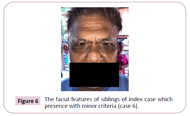
Figure 6: The facial features of siblings of index case which presence with minor criteria (case 6).
Case 7
The same condition also presence with a 28 years old nephew. He also presence with premature greying of the hair (below 30 years), synophris and broad high nasal root (Figure 7).
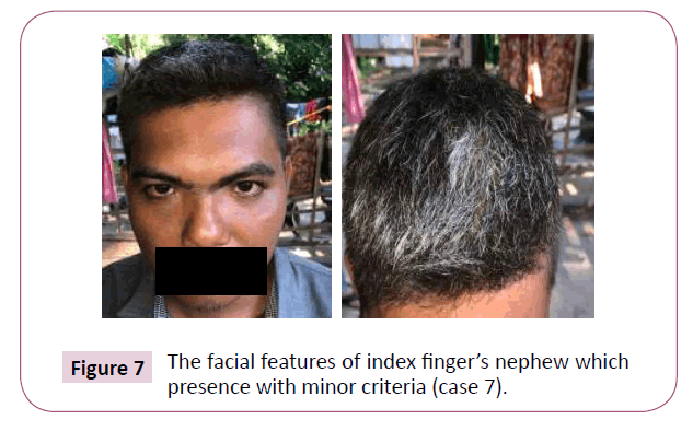
Figure 7: The facial features of index finger’s nephew which presence with minor criteria (case 7).
Case 8
A 4 years old nephew presence with the major criteria of waardenberg. The boy presence with heterochromia of iris and broad nasal bridge.
There were no delayed speech , while auditory examination couldn’t be done due to patient’s cooperation (Figure 8).
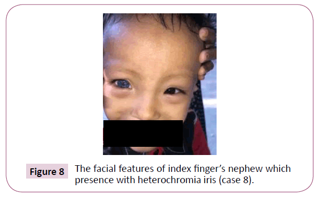
Figure 8: The facial features of index finger’s nephew which presence with heterochromia iris (case 8).
Case 9
4 years old nephew also presence with the major criteria of waardenberg. The boy presence with heterochromia of iris and broad nasal bridge. There were no delayed speech and no sensory neural deafness (Figure 9).
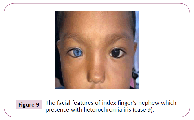
Figure 9: The facial features of index finger’s nephew which presence with heterochromia iris (case 9).
Case 10
A Niece also presence with bilateral bright irides. She presence with broad nasal bridge, hypopigmentation of the skin, have been assessed with ENT and diagnosed with sensory neural deafness (Figure 10).
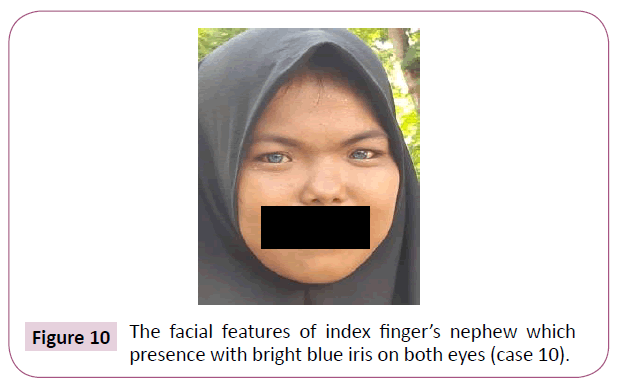
Figure 10: The facial features of index finger’s nephew which presence with bright blue iris on both eyes (case 10).
Discussion
Based on the findings, 10 subjects of in the family presents with criteria of wardenberg syndrome. We found that 4 subjects presence with bright blue iris, while 3 subjects presence with heterochromia iris. Partial or complete heterochromia can be presence in 21-21% of the patients with waardenberg syndrome [6-12].
Among 10 subjects, all of them also indicates widening of the nasal bridge and hypertelorism. This clinical feature is a crucial finding to differentiate the type 1 and type 2 waardenberg syndrome [9-13].
5 subjects was born with poliosis. In previous study, 8.5-50% patient with waardenberg syndrome can be presence with cutaneous pigmentary defect (Figure 11) [12-14].
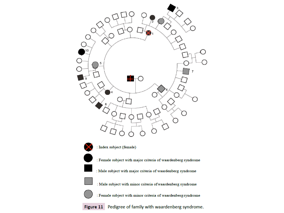
Figure 11: Pedigree of family with waardenberg syndrome.
Sensory neural deafness can be found among 2 patients, where statistically 2% person of congenital deafness is caused by waardenberg syndrome. In total, 69% of type 1 waardenberg syndrome can be presence with sensorineural deafness, while 87% of type 2 waardenberg syndrome can be presence with sensorineural deafness (Table 1) [12,15,16].
| Major Criteria |
Minor Critera |
| Congenital sensorineural deafness |
Congenital leukoderma, several areas of hypo pigmented skin |
| Pigmentary disorder of Iris, complete heterochromia iridis, partial or segmental heterochromia iris/ hypoplastic blue iris |
Pigmentary disturbance of iris, complete heterochromia iridis partial or segmental |
| White forelock, hair hypo pigmentation (poliosis) |
Broad and high nasal root |
| Dystopia Cantorum |
Hypoplasia of alae nasi |
| A?ected First Degree Relative , diagnosed with waardenberg syndrome |
Prematur graying of hair within age<30 years |
Table 1 Pedigree of family with waardenberg syndrome.
In the family within 4 generations, there were no consanguineous marriage. The family members whose presence with waardenberg syndrome , major or minor criteria, inherit generations with members presence with major or minor signs of waardenberg syndromes. Hence, the family without the signs didn’t phenotypically inherit the syndromes. Hence, the examination cannot be completed to posterior segment due to the disapproval of patients. This condition shows the waardenberg syndrome in this family can be considered autosomal dominant inheritance [13-16].
Kumar S in 2012 stated that Waardenberg is further divided into 4 types based on the genes; PAXIII being responsible for the presentation of types I and III; MITF responsible for the presentation of Waardenberg syndrome type II; and EDNRB and EDN3 responsible on the presentation of waardenberg syndrome type IV. Based on the assessment that already been done, the condition of syndrome in the family can be classified as type I waardenberg syndrome. Hence, the subjects in the family need to undergo further laboratory examination to genetically define the syndrome (Table 2) [2,6-8].
| NO |
Pigmentary disorder of Iris |
Pigmentary disturbance of iris |
Broad and high nasal root |
Dystopia Cantorum |
A?ected First Degree Relative |
Congenital leukoderma |
Poliosis |
Prematur graying of hair (<30y.o) |
Congenital sensorine uraldeafness |
Syno phris |
| 1 |
+ |
|
|
+ |
+ |
+ |
+ |
|
|
|
| 2 |
+ |
|
|
+ |
+ |
|
+ |
|
+ |
|
| 4 |
|
+ |
+ |
|
+ |
|
|
|
|
|
| 3 |
+ |
|
|
+ |
+ |
+ |
|
|
|
|
| 5 |
|
|
|
+ |
+ |
+ |
+ |
+ |
|
|
| 6 |
|
|
|
+ |
+ |
+ |
+ |
+ |
|
+ |
| 7 |
|
|
|
+ |
+ |
+ |
+ |
+ |
|
+ |
| 8 |
|
+ |
+ |
|
+ |
|
|
|
|
|
| 9 |
|
+ |
+ |
|
+ |
|
|
|
|
|
| 10 |
+ |
|
|
+ |
+ |
|
|
|
+ |
|
Table 3 The clinical features of subjects with waardenberg syndrome.
Conclusion
The large number of family which can be examined conclude that this is the first documented case with a family history suggesting autosomal dominance inheritance with a high degree of penetrance. The presence of major and or minor signs of waardenberg syndrome is a crucial key to the inheritance of the syndrome.
During the writings of this study, the genetic examination hasn’t been done and the genetic counselling couldn’t have done. Due to our limitation and geographical barrier, we couldn’t assess the whole family members.
35349
References
- Waardenburg PJ (1951) A new syndrome combining developmental anomalies of the eyelids, eyebrows and nose root with pigmentary defects of the iris and head hair and with congenital deafness. Am J Hum Genet. 3:195-253.
- Kumar S, Rao K (2012) Waardenberg syndrome: A rare genetic disorder, a report of two cases. Indian J Hum Genet. 18:254-255.
- Liu XZ, Newton VE, Read AP (1995) Waardenberg syndrome type II: Phenotypic findings and diagnostic criteria. A J Med Genet. 55:95-100.
- Hageman MJ, Delleman JW (1997) Heterogeneity in waardenberg syndrome. A J Hum Genet. 29:468-485.
- Shields C, Nickerson SJ, Al-Dahmash S, Shields J (2013) Waardenberg syndrome: Iris and choroidal hypopigmentation findings on anterior and posterior segment imaging. JAMA Ophthalmol. 131:1167-1173.
- Vichare LC, Bhargava CN (2013) Waardenberg Syndrome: A rare case with bilateral congenital cataract, an unusual entity. Med J Armed Forces India. 69:172-174.
- Faivre L, Vekemans M (2005) Waardenberg Syndrome type 1. Orphanet Encyclopedia.
- Read AP, Newton VE (1997) Waardenberg Syndrome. J Med Genet 34:656-665.
- Wadhwani M, Gupta YK, Gangwani K (2015) Waardenberg shah syndrome : a rare case from india. Oman Journal of Ophthalmology 8:74-75.
- Moser E, Eidenbock AM. Frisch H, Read AP (2001) Waardenberg type 2 in a turkish family: implications of the importance of the patters of Fundus Pigmentation. British Journal of Ophthalmology 85:1384-1386.
- Shiuey Y (1997) 29 years old man with visual symptoms and headaches. Digital Journal of Ophthalmology 3:14.
- Work TM, Shihab ZM, Young RSL, Price J (1986) Pigment distribution in waardenberg’s syndrome: A new hypothesis. Graefes Arch Clin Exp Ophthalmol. 224:487-492.
- Onabolu OO (2002) Waardenberg syndrome in a nigerian family. Nigerian Journal of Ophthalmology 10:32-34.
- Torgerson (2014) Genetic of primary immune deficiencies, stiehms immune deficiency.
- Tukenmez DG, Guldehan A, Kıvanc IK (2011) Waardenberg syndrome type 1 : a case report. Dermatology online journal. 17:3.
- Kassem LH, Ahmado MF, Alganameh MS (2018) A rare case of 7 siblings with waardenberg syndrome : a case report. Journal of medical case reports. 12:192.


