Keywords
Acetabulum; Femoral head; Anteversion; Abduction angle; Neck-shaft angle; Superior axial Coverage angle; Anterior axial coverage angle; Calculation formula
Background
The stabilization of hip joint was maintained by the bony acetabulum covering femoral head, articular cartilage, joint capsule, and surrounding soft tissue. Acetabular and femoral head spatial orientation angles include acetabular abduction angle, anterversion and femoral neck-shaft angle, and anteversion. The exact match of these spatial orientation angles plays a vital role in maintaining the normal range of motion and joint stability. Without this exact match will result in hip dysplasia [1,2], lead to hip instability or femoral acetabular impingement, cause hip wear, followed by hip osteoarthritis [3-9]. The current study on hip phase parameters mainly concentrated on the acetabulum supported femoral head. The femoral head and neck phase parameters were not taken into account, because they did not reflect the intrinsic relationship between acetabular abduction angle and anteversion, and femoral neck-shaft angle and anteversion. This study was designed to establish 3D coordinate systems concerning the anterior and superior axial coverage angle of acetabulum to femoral head, which display the intrinsic relationship of the spatial orientation angles between the acetabulum and femoral head. Eventually, the mathematical formulas were built and the influence factors were evaluated.
Methods
The mathematical calculation of anterior and superior axial coverage angle of acetabulum to femoral head in the absence of acetabulum and femoral neck anteversion.
The 3D coordinate system was established by setting O as the hip rotation center, X axis as the body coronal axis, Y-axis as the body vertical axis, Z-axis as the body sagittal axis (Figure 1). In this system, AB served as the femoral shaft axis, DA as femoral head-neck axis, and Y-axis parallel to AB axis. The angle b formed by OC and Y axis was acetabular abduction angle and ∠OAB was the femoral neck-shaft angle indicated by J. ∠COD was formed by segment OD (femoral head-neck axis) and segment OC. Segment OC was formed by the intersection of the plane where Y-axis and femoral head-neck axis lie in and the acetabular exit plane. ∠COD was named as superior axial coverage angle of acetabulum to femoral head. ∠EOD was formed by segment OD (femoral head-neck axis) and segment OE. Segment OE was formed by the intersection of the plane where Z-axis and femoral head-neck axis lie in and the acetabular exit plane. ∠EOD was named as anterior axial coverage angle of acetabulum to femoral head.
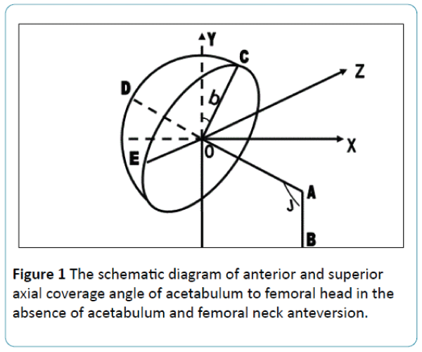
Figure 1: The schematic diagram of anterior and superior axial coverage angle of acetabulum to femoral head in the absence of acetabulum and femoral neck anteversion.
Without acetabular and femoral neck anteversion, the plane femoral head-neck axis and Y-axis lie in and the plane femoral head-neck axis and Z-axis lie in were perpendicular to the plane of the acetabular exit. So in this case the superior axial coverage angle of acetabulum to femoral head equals to the abduction angle plus the supplementary angle of femoral neck-shaft angle. It can be expressed as ∠COD = b + 180-J.
The mathematical calculation of superior axial coverage angle of acetabulum to femoral head in the presence of acetabulum and femoral neck anteversion.
The 3D coordinate system was built by setting the hip rotation center as O, X axis as the body coronal axis, Y-axis as the body vertical axis, Z-axis as the body sagittal axis (Figure 2). In this system, AB was the femoral shaft axis, DA as femoral head-neck axis and Y-axis parallel to AB axis. ∠OAB was femoral neck-shaft angle indicated by J and the angle b formed by OC and Y axis was acetabular abduction angle. ∠EOD was formed by segment OD (femoral head-neck axis) and segment OE. Segment OE was formed by the intersection of the plane where Y-axis and femoral head-neck axis lie in and the acetabular exit plane. ∠EOD was named as superior axial coverage angle of acetabulum to femoral head.
The plane OC and Z-axis lie in was perpendicular to the plane of acetabular exit and Y-axis was perpendicular to the plane EFG. Based on the 3D-system (Figure 2), the superior axial coverage angle of acetabulum to femoral head can be calculated using this equation: ∠EOD = ∠FOE + 180-J. ∠EFG equals to femoral neck anteversion plus acetabular anteversion A. ∠FOC' was acetabular abduction angle indicated by b. It was a projection angle in the coronal plane formed by ∠FOC forward A around the Y axis. So ∠FOC = arctg (tgb/cosA). ∠FOC was a projection angle of ∠FOE in the plane COF formed by ∠FOE forward A+a around the Y axis. So ∠FOE = arctg {tgb/[cosAcos (A + a)]}. Therefore, the superior axial coverage angle of acetabulum to femoral head ∠EOD = ∠FOE + 180-J = arctg {tgb/[cosAcos (A + a)]} + 180-J.
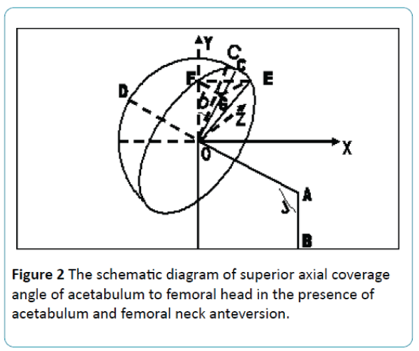
Figure 2: The schematic diagram of superior axial coverage angle of acetabulum to femoral head in the presence of acetabulum and femoral neck anteversion.
The mathematical calculation of anterior axial coverage angle of acetabulum to femoral head in the presence of acetabulum and femoral neck anteversion
As shown in Figure 3, the 3D coordinate system was built by setting O as the hip rotation center, X axis as coronal axis of the body, Y-axis as the body vertical axis, Z-axis as the body sagittal axis. In this system, AB was the femoral shaft axis, DA as femoral head-neck axis, and Y-axis parallel to AB. ∠OAB was the femoral neck-shaft angle indicated by J and the plane COD was perpendicular to the acetabular exit plane in the absence of acetabular and femoral neck anteversion. ∠EOD was formed by segment OD (femoral head-neck axis) and segment OE. Segment OE was formed by the intersection of the plane where Z-axis and femoral head-neck axis lie in and the acetabular exit plane. ∠EOD was named as anterior axial coverage angle of acetabulum to femoral head. Y-axis was perpendicular to the plane FOG. ∠HOC' was acetabular abduction angle b.
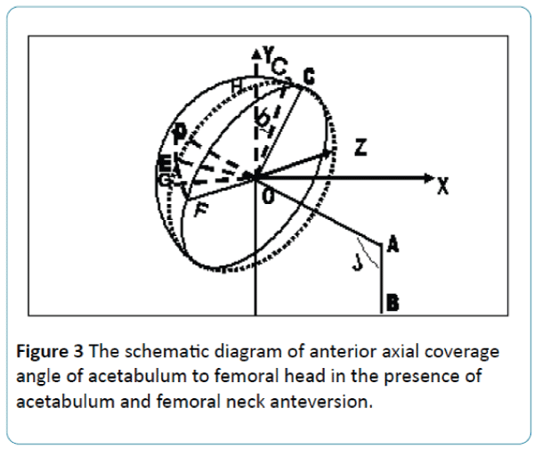
Figure 3: The schematic diagram of anterior axial coverage angle of acetabulum to femoral head in the presence of acetabulum and femoral neck anteversion.
Based on the system, the anterior axial coverage angle of acetabulum to femoral head can be calculated by ∠GOD =∠FOD-∠FOE = 90-∠FOE. ∠GOF equals to femoral neck anteversion plus acetabular anteversion A. ∠HOC' was acetabular abduction angle b, which was a projection angle in the coronal plane formed by ∠HOC forward A+a around the Y axis. So ∠HOC = arctg [tgb / (cosA + a)]. ∠GOF was A + a, which was a projection angle in the cross section formed by ∠FOE rotate arctg [tgb / (cosA + a)] - (J-90) around the Z axis. So ∠FOE = arctg [tg (A + a) / cos {arctg [tgb / (cosA + a)] - (J-90)}]. Thus, the anterior axial coverage angle of acetabulum to femoral head can be expressed as ∠GOD = 90- arctg [tg (A + a) / cos {arctg [tgb / (cosA + a)] - (J-90)}].
Results
In the absence of acetabular and femoral neck anteversion, the superior axial coverage angle of acetabulum to femoral head equals to the abduction angle plus the supplementary angle of femoral neck-shaft angle. It can be expressed as: ∠COD = b + 180-J (Figure 1). It was observed that the superior axial coverage angle of acetabulum to femoral head was positively correlated with acetabulum abduction angle and negatively with femoral neck-shaft angle. The anterior axial coverage angle of acetabulum to femoral head equals to 90 degrees. However, the anterior axial coverage angle of acetabulum to femoral head was not correlated with acetabular abduction angle and femoral neck-shaft angle.
In the presence of acetabulum and femoral neck anteversion, the superior axial coverage angle of acetabulum to femoral head can be expressed as: arctg {tgb / [cosAcos (A + a)]} + 180-J (Figure 2) and the anterior axial coverage angle of acetabulum to femoral head can be calculated using the following formula: 90- arctg [tg (A + a) / cos {arctg [tgb / (cosA + a)] - (J-90) }] (Figure 3). According to the principle known trigonometric functions, the superior axial coverage angle of acetabulum to femoral head was positively correlated with acetabular abduction angle, negatively with femoral neck-shaft angle, positively with acetabular and femoral neck anteversion. The anterior axial coverage angle of acetabulum to femoral head was negatively correlated with acetabular abduction angle, positively with femoral neck-shaft angle, negatively with acetabular and femoral neck anteversion.
Discussion
The coverage acetabular to the femoral head was essential for the stability of the hip joint. The radiographic parameters for characterizing it include anterior acetabutar sector angle(AASA), posterior acetabutar sector angle(PASA), horizontal acetabutar sector angle(HASA), and the center-edge angle(CE).
AASA was formed by the connecting line of two centers of the femoral head and the connecting line of acetabulum front edge and femoral head center. PASA was formed by the connecting line of two centers of the femoral head and the connecting line of the rear edge of the acetabulum and femoral head center. HASA is the combination of AASA and PASA. AASA, PASA, and HASA can provide anterior and posterior containment situation of acetabulum to femoral head [10]. Nevertheless, it was only impacted by the acetabular anteversion, not by acetabular abduction angle, femoral neck-shaft angle, and anteversion.
CE angle was the center-edge angle formed by the connecting line of the femoral head center and the outersuperior edge of the acetabulum and the vertical line through the femoral head center. It was the reflection of upper containment of acetabulum to femoral head [11], which only correlated with the acetabular abduction angle and anteversion. It was not relevant with femoral neck-shaft angle and anteversion. CE angle was not a true reflection of containment of acetabulum to femoral head.
In current study, the anterior and superior axial coverage angles of acetabulum to femoral head were determined by acetabular abduction angle, acetabular anteversion, femoral neck-shaft angle, and femoral neck anteversion. It not only locked the spatial alignment of acetabulum to femoral head, but also was a true reflection of containment situation of the acetabulum to femoral head. There was a close inherent relationship between acetabular abduction angle, acetabular anteversion, femoral neck-shaft angle, and femoral neck anteversion.
The anterior axial coverage angle of acetabulum to femoral head equals to 90°.
In the presence of the acetabular and femoral neck anteversion, the superior axial coverage angle of acetabulum to femoral head equals to arctg {tgb / [cosAcos (A + a)]} + 180- J. It was positively correlated with acetabular abduction angle, negatively with femoral neck-shaft angle, positively with acetabular and femoral neck anteversion. The anterior axial coverage angle of acetabulum to femoral head equals to 90-arctg [tg (A + a) / cos {arctg [tgb / (cosA + a)] - (J-90)}]. It was negatively correlated with acetabular abduction angle, positively with femoral neck-shaft angle, negatively with acetabulum and femoral neck anteversion.
Although the present study was based on semi-circular acetabulum, even acetabulum in the weak semi-circle, the anterior and superior axial coverage angle of acetabulum to femoral head can also similarly lock the spatial alignment of acetabulum to femoral head. In short, there was a close inner relationship of spatial angles between the acetabulum and femoral head, which exactly maintain the anterior and superior axial coverage of the acetabulum to the femoral head, maintain hip stability and meet hip normal range of motion. At present, direct imaging measurement of the anterior and superior axial coverage angle of acetabulum to femoral head is difficult. The imaging measurement of the acetabular abduction angle, anteversion, femoral neck anteversion and neck-shaft angle has been established. Consequently, the anterior and superior axial coverage angle of acetabulum to femoral head could be readily calculated using the mathematical formulas built in the current study. For example, based on the normal range of acetabular abduction angle (45° ± 10°) [12-16], acetabular anteversion (15° ± 10°) [12-15,17-19], femoral neck-shaft angle (110°-140°) [20], and the femoral neck anteversion (12°-15°) [21,22] in the previous studies, the normal ranges of the superior and anterior axial coverage angle of acetabulum to femoral head can be calculated out by the mathematical formulas being 76.32°~134.07° and 41.62°~72.91°, respectively.
Conclusions
In summary, the formula was built based on a 3D coordinate system established in this study. The formula reflects the inner relationship of spatial orientation angle of acetabulum to femoral head, which will offer a simple, novel and efficient mathematical method to calculate the anterior and superior axial coverage angle.
Competing interests
The authors declare that they have no competing interests.
9762
References
- Crowe JF, Mani VJ, Ranawat CS (1979) Total hip replacement in congenital dislocation and dysplasia of the hip. J Bone Joint Surg Am 61:15-23.
- Garvin KL, Bowen MK, Salvati EA, Ranawat CS ( 1991) Long-term results of total hip arthroplasty in congenital dislocation and dysplasia of the hip. A follow-up note. J Bone Joint Surg Am 73:1348-1354.
- Eijer H, Myers SR, Ganz R (2001)Anteriorfemoroacetabular impingement after femoral neck fractures. J Orthop Trauma 15: 475-481.
- Goodman DA, Feighan JE, Smith AD, Latimer B, Buly RL(1997) Subclinical slipped capital femoral epiphysis: relationship to osteoarthrosis of the hip. J Bone Joint Surg Am 79:1489-1497.
- Snow SW, Keret D, Scarangella S, Bowen J (1993) Anterior impingement of the femoral head: a late phenomenon of Legg-Calvé-Perthes’ disease. J PediatrOrthop13: 286-289.
- Myers SR, Eijer H, Ganz R (1999)Anteriorfemoroacetabular impingement after periacetabular osteotomy. ClinOrthopRelat Res 363: 93-99.
- Reynolds D, Lucas J, Klaue K (1999) Retroversion of the acetabulum: a cause of hip pain. J Bone Joint Surg Br8: 281-288.
- Ziebarth K, BalakumarJ, Domayer S, Kim YJ, Millis MB (2011) Bernese periacetabular osteotomy in males: is there an increased risk of femoroacetabular impingement (FAI) after Bernese periacetabular osteotomy? J ClinOrthopRelat Res 469:447-453.
- Imai H, Kamada T, Takeba J, Shiraishi Y, Mashima N, et al. (2014)Anterior coverage after eccentric rotational acetabular osteotomy for the treatment of developmental dysplasia of the hip. J OrthopSci 19: 762-769.
- Anda S, Svenningsen S, Dale LG (1986)Theacetabular sector angle of the adult hip determined by computed tomography. ActaRadiolDiagn (Stockh) 27: 443-447.
- Delaunay S, Dussault RG, Kaplan PA (1997) Radiographic measurements of dysplastic adult hips. Skeletal Radiol 26: 75-81.
- Lima DD, Urquhart AG, Buehler KO, Walker RH, Colwell CW (2000) The effect of the orientation of the acetabular and femoral components on the range of motion of the hip at different head neck ratios. J Bone Joint Surg 82: 315-321.
- Kummer F, Shah S, Iyer S, DiCesare P (1999) The effect of acetabular cup orientations on limiting hip rotation. J Arthroplasty 14: 509-513.
- Lewinnek GE, Lewis JL, Tarr R, Compere CL, Zimmerman JR (1978)Dislocations after total hip replacement arthroplasties. J Bone JointSurg 60: 217-220.
- Wentzensen A, Zheng G, Vock B, Langlotz U,Korber J, et al. (2003) Image based cup navigation. IntOrthop 27: 43-46.
- Hirakawa K, Mitsugi N, Koshino T, Saito T, Hirasawa Y, et al. (2001) Effect of acetabular cup position and orientation in cemented total hip arthroplasty. ClinOrthopRelat Res 388:135-142.
- Rittmeister M, Callitsis C (2006) Factors Influencing Cup Orientation in 500 Consecutive Total Hip Replacements. ClinOrthopRelat Res 445:192-196.
- Charnley J (1970) Total hip replacement by low friction arthroplasty. Clin OrthopRelat Res 72: 7-21.
- Coventry MB (1985)Late dislocations in patients with Charnley total hip arthroplasty. J Bone Joint Surg 67: 832-841.
- HoushanLv(1998)Artificial joint surgery. First Edition. Beijing: Science and Technology Press 9.
- Yi cong WANG editor (2001) Bone and joint injury. Beijing: People's Medical Publishing House842.
- Ma RF, Liu ShL, Xu J(2002) The measurement of proximal femur and its clinical significance. Clinical J Orthopaedics 5: 164-166.

