Research Article - (2022) Volume 10, Issue 8
A Split Mouth Clinical Trial to Compare Between Two Different Methods for Gingival Depigmentation-Scalpel and Diode Laser
Lashika Tambe*
Department of Periodontics, Nair Hospital Dental College, Mumbai, India
*Correspondence:
Lashika Tambe,
Department of Periodontics, Nair Hospital Dental College, Mumbai,
India,
Email:
Received: 07-Jul-2022, Manuscript No. IPACLR-22-3252;
Editor assigned: 08-Jul-2022, Pre QC No. IPACLR-22-3252(PQ);
Reviewed: 22-Jul-2022, QC No. IPACLR-22-3252;
Revised: 26-Jul-2022, Manuscript No. IPACLR-22-3252(R);
Published:
02-Aug-2022, DOI: 10.36648/2386- 5180.22.10.426
Abstract
Background: With the growing awareness about esthetics, gingival hyperpigmentation is one of the most common chief complain among patients now a days. There are various non-surgical and surgical methods introduced recently for gingival depigmentation. Laser is one of the most advanced technology used in dentistry for several surgical procedures. However, its expensive and technique sensitive. Thus it is important to justify the use of Laser for surgical procedures like Gingival depigmentation.
Aim: This case study presents an original research done to compare the efficacy, patient preference and satisfaction between Laser and Scalpel for gingival depigmenation.
Material and methods: A split mouth study was done among 20 patients to compare the efficacy of Diode Laser (Ezlase 940 nm) and scalpel. Clinical parameters were Dummet Oral Pigmentation Index (DOPI) and visual analog scale. Patients were recalled after regular intervals-once every week for initial 1 month, 3 month and 6 month.
Result: Non significant results between all the clinical parameters between both the techniques.
Conclusion: Both, Laser and scalpel provide similar clinical outcome for gingival depigmenation.
Keywords
Gingival hyperpigmentation; Melanin; Diode laser; Scalpel
Introduction
An esthetic smile is a perfect balance between white and pink components of the oral cavity. The colour of the gingiva is determined by various factors like number and size of underlying blood vessels, epithelial thickness, degree of keratinization, and residing pigments within the gingiva. Unesthetic appearances may be due to various factors one of which is pigmentation of gingiva. This may even have psychological impact on the patient. Such problem is aggravated in the presence of high smile line [1]. Gingival hyperpigmentation can be defined as a darker gingival colour beyond what is normally expected [2]. Various by-products of the physiological process such as melanin, melanoid, carotene, oxyhaemoglobin, reduced haemoglobin, bilirubin and iron contribute to the pigmentation [3]. Melanin is a brown pigment synthesized in the cytoplasm of melanocytes. Excessive melanin deposition in the basal and suprabasal cell layers of the gingival epithelium creates pigmented areas in gums [4].
Gingival depigmentation is a periodontal plastic surgery procedure, and variety of depigmentation techniques have been described in the literature. Most commonly used methods are surgical methods which include conventional techniques like scalpel, free gingival grafting and electro surgery [5]. Laser is an advanced and latest technology used for many periodontal surgical procedures. However, it is highly expensive as well as technique sensitive. Hence, the goal is to justify the use of an expensive, technique sensitive procedure like Laser for gingival depigmentation. The aim of this study is to compare and provide clinical evidence between efficacy of two different gingival depigmentation techniques: Scalpel and laser.
Materials and Methods
This was a Randomized controlled split mouth trial. A total of 20 patients with age range between 20 and 40 years were selected for the study.
Inclusion criteria
• Subjects suffering from bilateral gingival hyperpigmentation.
• Patients with primary esthetic concern in the anterior teeth region.
• Subjects were periodontally healthy were included in the study.
Exclusion criteria
• Patients with a history of systemic diseases like Diabetes or Hypertension.
• Patients with acute swelling or pain in the anterior teeth region.
• Patients with a history of smoking or tobacco chewing.
All the patients were explained about the surgical procedure and its possible complications. The subjects were voluntarily asked to sign an informed consent in order to participate in the study. The Maxillary arch was divided into two segments:
Segment I: Right Side from first premolar to right central incisors used for Laser depigmentation.
Segment II: Left Side from first premolar to left central incisors used for Scalpel depigmentation.
Procedure
Each patient underwent oral prophylaxis with ultrasonic scaling, one week before therapy. After 7 days the patient was scheduled for the gingival depigmentation procedure. The baseline pigmentation was measured prior to the procedure. The research included the following clinical evaluation parameters:
Dummet oral pigmentation index by Dummet and Gupta (1964): The index is used to score criteria gingival pigmentation and intensity of gingival hyperpigmentation. Scoring criteria are as follows: No clinical pigmentation (pink gingiva), Mild clinical pigmentation (mild brown colour), Moderate clinical gingival of pigmentation (medium brown or mixed pink and brown colour) Heavy clinical pigmentation (deep brown or bluish black colour).
Visual analogue scale for pain: The VAS was used to describe measure the intensity of pain experienced during and after treatment. The VAS consists of a horizontal line 100 mm long, anchored at the left end by the descriptor “no pain” and at the right end by ‘‘unbearable pain”. The patient placed a mark to coincide with the level of pain.
The VAS score was recorded during intraoperative treatment phase, and all the patients were recalled after 1st day and at 7th day for pain assessment [6].
Surgical procedure
Depigmentation with diode laser: Local infiltration with anesthetic solution Lignox® (2% lignocaine with 1:200,000 adrenaline) was given. As per the LASER safety rules, special eye glasses were worn by the patient and the staff. Highly reflective instruments or instruments with mirrored surfaces were avoided. The properly initiated tip of the diode LASER unit (EZLASE, wavelength 940 nm) angled at an external bevel of 45° and at energy settings of 4-4.5 W Continuous Wave (CW) was used with small brush like strokes back and forth with gradual progression to remove the tissue. Normal saline-soaked cotton or gauze was used to remove epithelial remnant [7].
Depigmentation with scalpel: Local infiltration with anesthetic solution Lignox® (2% lignocaine with 1:200,000 adrenaline) was given.15 no. surgical blade was used for this procedure. Partial thicknes incision flap was used to peel off the pigmented gingival tissues. The bleeding was controlled by compression pack.
Results
There is no significant difference between the baseline values for both the techniques. There was absolutely no pigmentation at 1, 2, 3 week and 1 month duration. There was no significant difference in the clinical outcomes of both the techniques after 6 months(Tables 1 and 2) [8].
| Time |
Laser, mean ± SD |
Scalpel, mean ± SD |
P value |
| 1 week |
2.75 ± 0.14 |
2.70 ± 0.14 |
0.75 |
| 2 week |
0.00 ± 0.00 |
0.00 ± 0.00 |
NA |
| 3 week |
0.00 ± 0.00 |
0.00 ± 0.00 |
NA |
| 1 month |
0.00 ± 0.00 |
0.00 ± 0.00 |
NA |
| 3 month |
0.15 ± 0.11 |
0.21 ± 0.17 |
0.683 |
| 6 month |
0.20 ± 0.20 |
0.25 ± 0.18 |
0.675 |
| P value |
<0.001 |
<0.001 |
|
Table 1: Dummet oral pigmentation index.
| Time |
Laser, mean ± SD |
Scalpel, mean ± SD |
P value |
| Day 1 |
1.20 ± 0.20 |
1.00 ± 0.25 |
0.32 |
| Day 2 |
1.10 ± 0.15 |
1.00 ± 0.10 |
0.276 |
| Day 3 |
1.02 ± 0.12 |
0.90 ± 0.09 |
0.124 |
| Day 4 |
0.00 ± 0.00 |
0.00 ± 0.00 |
|
| Day 5 |
0.00 ± 0.00 |
0.00 ± 0.00 |
|
Table 2: Visual analog scale.
The patient complaint of mild to moderate pain and burning sensation on the sides of gingival depigmentation. Although the pain sensation was greater in sites where laser technique was used for gingival depigmentation, there was no statistically significant difference between both the sites. The pain completely reduced after 4th day with respect to both the techniques.
Bleeding during the procedure: There was statistically significant difference between the bleeding during the procedure (p value<0.05). There was no bleeding seen during gingival depigmentation performed by Laser as compared to Scalpel technique [9].
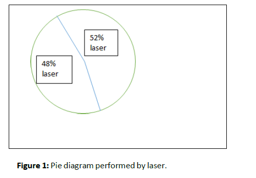
Patient satisfaction and preference: There was no significant difference between patient preference and satisfaction after 6 months between both the techniques (Figure 1).

Figure 1: Pie diagram performed by laser.
Discussion
Diode laser at 940 nm wavelength enables targeted radiation to soft tissue with excellent coagulation and cutting results. Depigmentation with the use of this laser is achievable due to its penetration into haemoglobin and melanin pigments [10]. At low powers, this laser is completely safe on dental hard tissues. Therefore it is applied as an excellent laser in soft tissue surgery. These Lasers are used for cutting and coagulation of gingiva and oral mucosa, as well as for soft tissue curettage or sulcular debridement with no adverse effect on root surfaces [11]. Diode laser hand piece is small and affordable. Lasers have been recognized as a reliable method for this kind of treatment with many reported advantages. Healing of laser wounds is slower than healing of scalpel wounds. It creates a sterile inflammatory reaction around the surgical area. But this approach needs expensive and sophisticated equipment that is not available commonly at all places. Hence it makes the treatment very expensive.
Risk of recurrence of gingival pigmentation exists in some cases of depigmentation treatment. Regimentation refers to the clinical appearance of melanin pigment following a period of clinical depigmentation [12]. The duration of repigmentation mentioned in literature remains controversial from one technique to other. The repigmentation depends on the methodology and the duration of follow-up. Furthermore, factors such as smoking, sun exposure, and genetic susceptibility also affect the repigmentation of gingiva. According to the migration theory, active melanocytes migrate from the adjacent pigmented tissues to the treated areas and result in recurrence of pigmentation [13]. In the current study, patients were followed up for three months and no case of recurrence was noted. This result may be due to the short duration of follow up. Difference in re-pigmentation time may be related to the technique of treatment and race of patients. It may also be related to presence of melanocytes in the areas adjacent to the surgical site.
Conclusion
The present study demonstrated that techniques, Diode Laser and Scalpel method used for gingival depigmentation provided same results as measured by Dummet Oral Pigmentation Index (DOPI) and Visual Analog Scale. Patient satisfaction was almost similar for both techniques. Recurrence of the gingival pigmentation was not seen in any of the cases. Within the limits of this study, we can conclude by saying that both scalpel and laser provide similar clinical outcomes. Although the amount of gingival bleeding is greater with the use of scalpel during gingival depigmentation, the patient satisfaction and preference is similar for both the techniques.
REFERENCES
- Malhotra S, Sharma N, Basavaraj P (2014) Gingival esthetics by depigmentation. J Periodontal Med Clin Pract 1: 79-84.
Google Scholar
- El-Shenawy H, Fahd A, Ellabban M, Dahaba M, Khalifa M (2017) Lasers for esthetic removal of gingival hyperpigmentation: A systematic review of randomized clinical trials. Int J Adv Res 5: 1238-1248.
Cross Ref
- Ozbayrak S, Dumlu A, Ercalik-Yalcinkaya S (2000) Treatment of melanin-pigmented gingiva and oral mucosa by CO2 laser. Oral Surg Oral Med Oral Pathol Oral Radiol Endodontol 90(1): 14-15.
Indexed at, Google Scholar, Cross Ref
- Lerner AB, Fitzpatrick TB (1950) Biochemistry of melanin formation. Physiol Rev 30(1): 91-126.
Indexed at, Google Scholar, Cross Ref
- Kumar S, Bhat GS, Bhat KM (2012) Development in techniques for gingival depigmentation-An update. Indian J Dent 3: 213-221.
Google Scholar, Cross Ref
- Shimada Y, Tai H, Tanaka A, Ikezawa-Suzuki I, Takagi K, et al. (2009) Effects of ascorbic acid on gingival melanin pigmentation in vitro and in vivo. J Periodontol 80: 317-323.
Indexed at, Google Scholar, Cross Ref
- Sheel V, Purwar P, Dixit J, Rai P (2015) Ancillary role of vitamin C in pink aesthetics. BMJ Case Rep 2015: bcr2014208559.
Indexed at, Google Scholar, Cross Ref
- Khalilian F, Nateghi Z, Janbakhsh N (2016) Gingival depigmentation using lasers: A literature review. Br J Med Med Res 12: 1-7.
Google Scholar
- Dummett CO, Gupta OP (1964) Estimating the epidemiology of oral pigmentation. J Natl Med Assoc 56(5): 419-420.
Indexed at,Cross Ref
- Huskisson EC (1974) Measurement of pain. Lancet 2(7889): 1127-1131.
Indexed at, Google Scholar, Cross Ref
- Gupta G (2011) Management of gingival hyperpigmentation by semiconductor diode laser. J Cutan Aesthet Surg 4: 208.
Indexed at, Google Scholar, Cross Ref
- Srivastava S, Shrivastava T, Dwivedi S, Yadav P (2014) Gingival melanin depigmentation: A review and case report. J Orofac Res 5: 50-54.
Google Scholar
- Kanakamedala AK, Geetha A, Ramakrishnan T, Emadi P (2010) Management of gingival hyperpigmentation by the surgical scalpel technique-report of three cases. J Clin Diagn Res 14: 2341-2346.
Google Scholar
Citation: Tambe L (2022) A Split Mouth Clinical Trial to Compare Between Two Different Methods for Gingival Depigmentation-Scalpel and Diode Laser. Ann Clin Lab Res Vol: 10 No: 8:426.


