Introduction
Acute stroke in a 26 year old patient is a rare finding. In such cases, physicians usually assume an underlying medical problem to account for the symptoms. In this case, the patient underwent an extensive workup revealing no outright cause. Then, one brain scan identified an incidental upper cervical spine lesion. This case discusses the initial patient work-up and a rare presentation of neurosarcoidosis.
Case
A 26 year old Hispanic male presented to the emergency room with complaints of sudden onset of right sided weakness. The prior day he was in his normal state of health without any motor or sensory deficits. Upon waking that morning, the patient noticed difficulty speaking and was unable to move the right side of his body. The patient denied any fevers, chills, neck pain, visual changes, seizures, headache, or nausea.
Three months prior to this ER visit, the patient had a prolonged stay at another hospital. At that time he presented with a two day history of an elevated temperature to 105ºF, occipital headache, neck pain and stiffness, dizziness, nausea and vomiting, and anorexia. When that admitting team pried further, they discovered that the patient was from Mexico and had only been in this country for five years. In addition, the patient stated that he had a non-productive cough for nine months and a vague history of an untreated positive PPD.
While hospitalized, the team suspected meningitis and proceeded with the proper work-up. The patient experienced one episode of hallucinations and complained of pain in his posterior thighs that was not associated with any weakness.
Several lumbar punctures performed were all consistent with chronic meningitis, showing lymphocyte predominance and low glucose levels. Due to these findings, history of an untreated positive PPD, and while cultures were growing, the patient began anti-tuberculosis RIPE regimen (Rifampin, Isoniazid, Pyrazinamide, Ethambutol, and Pyridex) and oral steroids.
Although all cultures remained negative (blood, fungal, AFB), the patient’s headache resolved. He also began tolerating oral feeds, and he became more alert and oriented. Therefore, the patient was discharged and was complaint with a medication regimen that included RIPE and oral prednisone.
On presentation at this hospital, physical examination portrayed an afebrile patient in no acute distress. Neurologic exam identified slurred speech, right facial droop, brisk lower extremity reflexes, and 1/5 strength in the upper and lower right extremities.
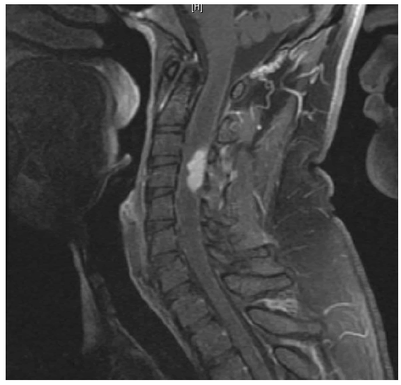
Imaging studies illustrated several findings. Initial brain MRI showed an acute stroke in the left coronal radiate, a 4-mm enhancement in the left temporal lobe without surrounding edema, and an incidental upper c-spine lesion likely within the spinal cord. For further clarification, a cervical MRI identified an enhancing, intra-medullary lesion in the right posterolateral cervical spinal cord extending from C3 to C4-5 level, containing a 5-mm cystic component with adjacent dorsal spinal cord edema or non-enhancing infiltrative changes extending from the skull base to T7 and adjacent syrinx. Brain MRA did not identify any vascular abnormalities.
This patient had a workup aimed to identify the cause of his stroke. All vasculitis investigations proved negative.
Due to limited services at the presenting hospital, the patient was transferred to a larger facility for further workup of the cervical spinal lesion.
On presentation, neurological exam showed right facial droop, sustained clonus of the right ankle, and increased muscle tone, decreased muscle strength, and increased reflexes in the right upper and lower extremities. The patient was able to stand and could walk only with assistance.
This patient underwent repeat imaging, cultures, and lumbar puncture, and since they yielded little additional information, the patient underwent both a brain and cervical spine biopsy. The brain biopsy from the left corona radiata showed findings consistent with a stroke and no evidence of neoplasm, demyelinated lesions, or infection. The pathology from the spinal biopsy identified lympho-plasmacytic infiltrates and non-caseating granulomas, consistent with neurosarcoidosis.
After these findings, RIPE therapy was discontinued and a slow prednisone taper was started. The patient continued to receive physical and occupational therapy, and he was transferred back to this hospital for discharge planning.
Discussion
The exact etiology of sarcoidosis is unknown, but proposed causes include infectious agents, occupational and environmental factors, genetic factors, and autoimmune disorders. [1] Sarcoid lesions can occur anywhere in the body but there is a predominance in the lungs, skin, and lymph nodes. [1] Neurosarcoidosis presents in less than five percent of individuals with sarcoidosis, and it usually occurs only after other systemic symptoms are found. [1] About 1% of sarcoidosis cases present with CNS problems alone. [2]
While sarcoidosis usually presents with remitting and relapsing episodes, neurosarcoidosis usually presents as a monophasic self-limiting illness. [1] Presentation will also vary in individuals depending on the location of the lesion with manifestations involving cranial nerves, parenchymal brain tissue, pituitaryhypothalamic axis, the spinal cord, and peripheral nerves. [1] Typical presentation include affects of the cranial nerves including facial palsy, visual loss, double vision, hearing loss, vertigo, swallowing problem, shoulder and tongue weakness. [2] Other presentations include grand mal seizures, meningitis, severe headaches caused increased ICP, and hydrocephalus. [2]
Although few cases have presented with symptoms of acute or subacute CNS ischemic events, the majority of these cases occurred in patients with known sarcoidosis including patients who were on treatment. Proposed mechanisms responsible for the cerebrovascular even include small vessel granulomatous vasculitis, large vessel inflammation leading to occlusion or stenosis, and embolism. [3,4]
As in this case, when systemic manifestations are absent, imaging and then biopsy are necessary to confirm the diagnosis. [1] Biopsy of lesions should identify non-caseating epithelioid-cell granulomas that over time should resolve or convert to hyaline connective tissue. [1] Although lumbar puncture may demonstrate an elevated protein level, pleiocytosis, and oligoclonal bands, about 30% of cases show no cerebral spinal fluid abnormality. [2] Various other tests (e.g. ACE level in CSF) have little added value in neurosarcoidosis. [2]
Mainstay treatment involves corticosteroids, immunosuppressants, and possible surgical excision of lesions. [1] Corticosteroids like prednisone are the main therapeutic agent in the management of neurosarcoidosis. [1] Small studies have found resistant cases to respond to immunosuppressants, including methotrexate, hydroxychloroquine, cyclophosphamide, pentoxifylline, thalidomide, and infliximab. [2] Radiotherapy and neurosurgical interventions are usually considered with obstruction or mass effect. [2] In one case of neurosarcoidosis that presented with acute stroke, surgeons performed left middle cerebral artery angioplasty with successful results. [5]
Since few cases of acute stroke caused by neurosarcoidosis have been reported, neurosarcoidosis as a cause of unexplained acute CNS ischemic event should be considered in a relatively young patient when other common etiologies have been ruled out. Diagnostic work-up should include routine laboratory studies, CSF analysis, imaging studies, and biopsy of abnormal lesions to develop a correct diagnosis in order to satisfactorily treat the patient.
2311
References
- Vinas FC, Rengachary S. Diagnosis and management of neurosarcoidosis. Journal of Clinical Neuroscience. 2001;8(6):505-513.
- Joseph FG, Scolding NJ. Sarcoidosis of the nervous system. Practical neurology. 2007; 7(4):234-244.
- Younger DS, Hays AP, Brust JC, Rowland LP. Granulomatous angiitis of the brain: an inflammatory reaction of diverse etiology. Arch Neurol. 1988; 45:514-518.
- Raske-Nielsen E, Harmsen A: Periangiitis as a manifestation of sarcoidosis of the brain: report of a case. J Nerv Ment Dis. 1962; 135:399-412.
- Brisman JL, Hinduja A, Mckinney JS, Gerthardstein B. Successful emergent angioplasty of neurosarcoid vasculitis presenting with strokes. Surgical Neuro. 2006; 66(4):402-404.

