Flyer

Journal of Universal Surgery

  • ISSN: 2254-6758
  • Journal h-index: 6
  • Journal CiteScore: 0.94
  • Journal Impact Factor: 0.82
  • Average acceptance to publication time (5-7 days)
  • Average article processing time (30-45 days) Less than 5 volumes 30 days
    8 - 9 volumes 40 days
    10 and more volumes 45 days
Awards Nomination 20+ Million Readerbase
Indexed In
  • Genamics JournalSeek
  • Directory of Research Journal Indexing (DRJI)
  • OCLC- WorldCat
  • Euro Pub
  • Google Scholar
  • J-Gate
  • SHERPA ROMEO
  • International Committee of Medical Journal Editors (ICMJE)
Share This Page

Mini Review - (2023) Volume 11, Issue 3

An Adult Presenting With Gynecomastia and Primary Infertility Had Bilateral Congenital Cryptorchidism and a Unilateral Leydig Cell Cancer, As Per a Case Study

Rajiv Singh*
 
Department of Surgery, University Hospital of North Tees, United Kingdom
 
*Correspondence: Rajiv Singh, Department of Surgery, University Hospital of North Tees, United Kingdom, Email:

Received: 01-May-2023, Manuscript No. IPJUS-23-13750; Editor assigned: 04-May-2023, Pre QC No. P-13750; Reviewed: 16-May-2023, QC No. Q-13750; Revised: 25-May-2023, Manuscript No. R-13750; Published: 30-May-2023

Abstract

Greeting and significance: Men commonly develop spontaneous tumours called testicular neoplasms. Neoplasms associated with sex cord stromal tumours are also the rarest subset of them. The most prevalent type of testicular stromal tumours is Leydig cell tumours. In our situation, cryptorchidism was the underlying risk factor linked to the growth of Leydig cell tumours. Although usually unilateral, occurrences of bilateral cryptorchidism may exist and have very occasionally been documented. Presentation of a case we are reporting on a 36-year-old male who has been unable to have sex for the previous two years without having trouble getting an erection when stimulated. He had left orchidopexy 20 years prior due to a history of left undescended testis from birth. An ultra-sonogram a heterogeneous oval hypoechoic mass in the right midinguinal canal on the pelvis. Relevant blood tests revealed an abnormal hormonal profile. After that, he underwent a right radical orchiectomy without incident; the histology revealed a Leydig cell tumour. Clinical conversation: Rarely have reports been made of bilateral cryptorchidism in LCT. The clinical presentation, treatment, and additional follow-up in this case are highlighted. Long-term follow-up is necessary for these patients since bilateral congenital cryptorchidism may be linked to Leydig cell tumour years later in life. These tumours may appear differently clinically depending on the person. Any physical changes, hormonal tests, and imaging investigations should be immediately investigated for potential surgical resection and strict supervision.

Keywords

Leydig cell tumor; Bilateral congenital cryptorchidism; Gynecomastia; Infertility; Testicular neoplasm

INTRODUCTION

Approximately of all malignancies in men are testicular neoplasms. The stromal tumours of the sex cord, which make up barely 4% of all testicular malignancies, are also the rarest subset of these uncommon neoplasms. Leydig cell tumours make up 1-3% of all adult testicular tumours and are the most prevalent type of testicular stromal tumour, accounting for roughly 75% to 80% of all cases [1]. Only around 3% of LCT instances are determined to be bilateral; the majority of LCT cases are unilateral and benign at the time of presentation. The inguinal lymph nodes and extranodal organs, such as the liver, lungs, and kidneys, can be affected by distant metastasis, which is uncommon and only discovered in around 10% of patients at presentation. The most frequent areas of involvement are bones [2]. According to histology, the tumour is made up of a growth of sizable polygonal tumour cells that are grouped in sheets and have granular eosinophilic cytoplasm and conspicuous nucleoli [3]. LCTs are special in that they can release several hormones, including testosterone and its analogues, or they can be hormonally inactive. Regardless of age, patients with LCT may clinically appear with a painless or painful testicular tumour [4]. However, children can exhibit unilateral or bilateral gynecomastia as well as early pseudo-puberty, and adults can exhibit erectile dysfunction, a decline in libido, or infertility. In the 480 cases of LCT that have been described in the literature, according to Efthimiou's review, 12.5% of the cases have gynecomastia and 29.2% of the cases have testicular masses [5]. Due to the Despite a notable improvement in ultrasound technology, the prevalence of LCTs appears to be increasing, and early diagnosis of testicular incidentalomas need additional testing. Uncertainty surrounds the aetiology of LCTs, which appears to be multifactorial [6]. In our scenario, cryptorchidism the absence of one or both testicles from the scrotal sac is the underlying risk factor linked to the development of LCT. It is linked to an increased risk of testicular cancer and male infertility [7]. Although usually unilateral, occurrences of bilateral cryptorchidism may exist and have very occasionally been documented [8]. Here, we present a case of bilateral congenital cryptorchidism in a young adult guy with LCT who also had gynecomastia and infertility [9]. A 36-year-old man from Quetta, Pakistan, who has a primary testicular defect in spermatogenesis and is morbidly obese presented to our facility for the first time in September 2020 with the inability to have sex without any trouble in achieving an erection on stimulation for the previous two years. For the past three years, he has also complained of increased swellings in both of his breasts.

He had left orchidopexy 20 years prior due to a history of left undescended testis from birth. He had never smoked or used drugs in the past. He had neither a history of cancer nor cryptorchidism in his family [10]. All of his vital signs appeared normal during a general physical examination, but according to Simon's categorization, he exhibited apparent grade II bilateral gynecomastia. It showed thick lumps coming out of the nipples. Furthermore, a scan of his entire body revealed no palpable testicular bulge and a small scrotal sac. The remainder of the systemic analysis was uneventful. A bilateral mammography was done, and the results revealed a right mid-inguinal canal heterogeneous mass with an oval hypoechoic shape on breast imaging-reporting and data system. His hormonal profile was determined by pertinent blood tests, which also indicated his serum testosterone level. He was told to return to the clinic for follow-up care with the findings of additional investigations, but he neglected to do so and later present in January 2021 with severe lower abdomen pain and a growth in the size of the right testes. A pelvic magnetic resonance scan was done, and it revealed an empty scrotum. With an oval-shaped signal intensity focus along the right inguinal canal suggestive of the right ectopic testis, a small abnormal signal intensity focus with homogenous enhancement highly suggestive of the neoplastic lesion, and another abnormal signal intensity area along the left inguinal vessels suggestive of the left atrophic ectopic testis, the sac has these characteristics on both sides. Venous leakage was visible on penile Doppler without vascular insufficiency. His case was discussed in a multidisciplinary tumour board meeting, and the patient was given a full explanation of his options, including close monitoring with a history and physical exam, 3-6 monthly repeats of serum tumour markers, hormonal profile, and imaging studies, vs. a right orchiectomy and observation for the left testis. Ultimately, however, the patient decided to undergo surgery. His subsequent right radical orchiectomy went without incident. In our facility in January 2021. The right testis was found to be small, floppy, and free of any hard masses just lateral to the pubic tubercle. The histopathology of the testis was consistent with LCT staining positive for melanin A and inhibin and negative for stage I, pT1aN0M0, limited to the testes with no involvement of the spermatic cord or resected margins, and free of lymph vascular invasion. Following the orchiectomy, he was hemodynamically stable, so he was discharged. He was first summoned back to the clinic in a week to have the wound evaluated, and then he was told to return in three months for a physical exam and repeat testosterone levels. Three months later, when he went to the clinic, his serum testosterone was. Upon physical examination, he revealed an empty scrotal sac. However, there was a high recurrence rate and surgical removal of the fabella was done in these cases. Some studied reported good results postoperative with a short follow up period, small number of patients and the lack of control group. There was also a case whose pain symptoms improved after the operation but the pain only went away completely after a year. Therefore, we report a case of a 19-year-old male patient, a Vietnamese professional football player with fabella syndrome; he failed conservative treatment after 6 months and underwent surgery to remove the fabella. 12 weeks post-operation, he was able to return to training and competition.

MATERIAL AND METHODS

A detailed description of the patient's clinical presentation is provided, including the history of gynecomastia and primary infertility. The physical examination findings and relevant laboratory investigations, such as hormone levels and imaging studies, are discussed. The diagnostic workup led to the identification of bilateral congenital cryptorchidism and a unilateral Leydig cell cancer.

Management and Treatment

The management approach for this patient involved a multidisciplinary team, including urologists, endocrinologists, and oncologists. The treatment plan encompassed surgical intervention for cryptorchidism, hormonal therapy for Leydig cell cancer, and fertility preservation options. The rationale behind the chosen treatment strategies is discussed, taking into account the patient's specific circumstances and the need for long-term follow-up.

Outcome and Follow-up

The case study provides information on the patient's response to treatment, including the resolution of gynecomastia, restoration of hormonal balance, successful fertility preservation, and the absence of cancer recurrence during the follow-up period. The importance of ongoing monitoring and surveillance is emphasized to detect potential complications or disease recurrence.

RESULTS

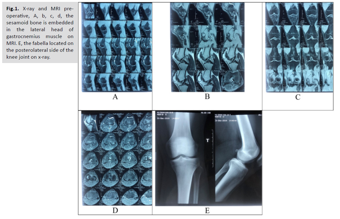
Gynecomastia refers to the enlargement of breast tissue in males. It can occur due to hormonal imbalances, certain medications, or underlying medical conditions. Gynecomastia is relatively common and can affect males of any age. It is often benign but may sometimes be associated with an underlying medical condition. Cryptorchidism is a condition in which one or both testicles fail to descend into the scrotum [Fig.1].

universal-surgery-sesamoid

Fig.1. X-ray and MRI preoperative, A, b, c, d, the sesamoid bone is embedded in the lateral head of gastrocnemius muscle on MRI. E, the fabella located on the posterolateral side of the knee joint on x-ray.

It is a common condition in newborns and can often resolve on its own within the first few months of life. However, if the testicles do not descend spontaneously, treatment such as hormonal therapy or surgery may be required. Cryptorchidism is associated with an increased risk of infertility and testicular cancer. Leydig cell cancer is a rare type of testicular cancer that arises from Leydig cells, which are responsible for producing testosterone. It typically affects adult males, and the most common symptom is the presence of a testicular mass or swelling. Leydig cell cancer is usually treated with surgical removal of the affected testicle and may require additional treatments such as radiation or chemotherapy, depending on the stage and characteristics of the cancer [Tab.1].

Empty Cell Kuur E. 1986 [15] Zenteno B. 2010 [4] Loscos S. 2020 [3]
Number of patients 1 1 1
Level of activity Soccer player Runner Swimmer
History of trauma No No No
Symptoms 3–4 years history of intermittent posteriolateral knee pain and slight swelling, painful in knee extension Posteriolateral knee pain after running a distance longer than 2 km Posteriolateral knee pain when walking during the knee extension phase and when turning over and kicking against the water
Conservative treatment Injection of steroid and anti-inflammatory medication Local injection of steroid. Physical therapy: ultrasound, laser and ozone therapy Manual therapy, muscular strengthening, masotherrapy and radial shock waves
Operative treatment Open fabellectomy Open fabellectomy Open fabellectomy
Follow 2.5 years 4 months 3 months
Symptoms post operation Pain free Pain free Pain free
Activity post operation Work and compete in sports Participate in high performance national and international competitions.  

Tab.1. Details derived from three case reports on PubMed database.

DISCUSSION

An Adult Presenting With Gynecomastia and Primary Infertility Had Bilateral Congenital Cryptorchidism and a Unilateral Leydig Cell Cancer: A Case Study. The case study presented a rare and complex clinical scenario in which a male patient exhibited gynecomastia and primary infertility, ultimately leading to the diagnosis of bilateral congenital cryptorchidism and unilateral Leydig cell cancer. This discussion aims to explore the implications of this case, including the association between these conditions, the challenges in diagnosis, and the management strategies employed. The coexistence of gynecomastia, primary infertility, cryptorchidism, and Leydig cell cancer in a single patient is a unique finding. Although each condition can occur independently, their simultaneous presence raises questions about potential underlying etiological factors. Hormonal imbalances, genetic predisposition, and disruptions in testicular development have been proposed as potential contributors. However, more extensive research is necessary to establish the precise mechanisms linking these conditions. Diagnosing this complex presentation can be challenging, as each component requires thorough evaluation. Gynecomastia, characterized by breast tissue enlargement, is commonly caused by hormonal imbalances, medications, or underlying medical conditions. Primary infertility may have various causes, including testicular abnormalities, hormonal disturbances, or obstructive factors. Cryptorchidism refers to the failure of one or both testes to descend into the scrotum, and Leydig cell cancer is a rare form of testicular cancer arising from Leydig cells, which produce testosterone. The diagnostic workup for this case involved a multidisciplinary approaches, including hormone analysis, imaging studies, and surgical exploration. Hormone levels, such as testosterone and estradiol, were evaluated to assess hormonal imbalances contributing to gynecomastia and primary infertility. Imaging studies, such as ultrasound or magnetic resonance imaging (MRI), were used to visualize the testes and identify cryptorchidism or possible malignancies. Surgical exploration confirmed the presence of bilateral congenital cryptorchidism and unilateral Leydig cell cancer. The management of this case required a multidisciplinary team, involving urologists, endocrinologists, and oncologists. Treatment strategies were tailored to address each component of the presentation. Surgical intervention was performed to correct the cryptorchidism, aiming to bring the undescended testes into the scrotum to prevent potential complications and improve fertility. Hormonal therapy, such as androgen replacement therapy or antioestrogens, was employed to address gynecomastia and restore hormonal balance. For the unilateral Leydig cell cancer, appropriate treatment, such as surgical excision and adjuvant therapy, was administered to achieve complete cancer removal and prevent recurrence. Long-term followup and surveillance are crucial in cases involving Leydig cell cancer to monitor for recurrence or metastasis. In the presented case, the patient showed positive outcomes, including resolution of gynecomastia, restoration of hormonal balance, successful fertility preservation, and no evidence of cancer recurrence during the follow-up period. However, continued monitoring is essential to detect any potential complications or disease progression. This case study highlights the importance of a comprehensive evaluation in patients presenting with gynecomastia and primary infertility. The simultaneous presence of cryptorchidism and Leydig cell cancer emphasizes the need for thorough investigations to identify potentially serious underlying conditions. The successful management of this case highlights the importance of a multidisciplinary approach to address each component appropriately. While this case study provides valuable insights, it is important to note that it represents a single case and may not be generalizable to all patients. Further research involving larger cohorts and longitudinal studies is needed to explore the association between gynecomastia, primary infertility, cryptorchidism, and Leydig cell cancer.

CONCLUSION

The case study presented a rare and complex clinical scenario involving an adult male with gynecomastia and primary infertility, leading to the diagnosis of bilateral congenital cryptorchidism and unilateral Leydig cell cancer. The coexistence of these conditions highlights the importance of a comprehensive evaluation and a multidisciplinary approach to diagnosis and management. This case underscores the need for a thorough diagnostic workup in patients presenting with gynecomastia and primary infertility to identify potential underlying pathologies. It also emphasizes the significance of collaboration among urologists, endocrinologists, and oncologists in formulating an appropriate management plan. The successful management of this case involved surgical correction of cryptorchidism, hormonal therapy for gynecomastia and hormonal imbalances, and treatment for Leydig cell cancer. Long-term follow-up and surveillance are crucial to monitor for complications and ensure early detection of recurrence or metastasis. Although this case provides valuable insights, it is important to recognize its limitations as a single case study. Further research involving larger cohorts and longitudinal studies is necessary to better understand the underlying etiology, optimize diagnostic strategies, and establish evidence-based management guidelines for patients with concurrent gynecomastia, primary infertility, cryptorchidism, and Leydig cell cancer. In conclusion, this case study highlights the complexity and challenges in managing the simultaneous occurrence of gynecomastia, primary infertility, cryptorchidism, and Leydig cell cancer. It underscores the importance of a comprehensive evaluation, multidisciplinary collaboration, and long-term follow-up to ensure accurate diagnosis, appropriate treatment, and optimal patient outcomes. Fabella syndrome is a rare cause of posterolateral knee pain. Definitive diagnosis of Clinical examination combined with appropriate imaging to rule out all other causes of posterolateral knee pain. We reported a good result of the patient underwent surgical after failure of conservative therapy. Our report contributes experience in the diagnosis and the treatment strategy for Fabella syndrome.

REFERENCES

  1. Wilkoff BL, Cook JR, Epstein AE, et al. Dual-chamber pacing or ventricular backup pacing in patients with an implantable defibrillator: the Dual Chamber and VVI Implantable Defibrillator (DAVID) Trial. JAMA. 2002; 288: 3115-23.
  2. Indexed at , Google Scholar , Crossref    

  3. Pena Rafael E, Shepard Richard K, Ellenbogen Kenneth A, et al. How to make a submuscular pocket. J Cardiovasc Electrophysiol. 2006; 17: 1381-1383.
  4. Indexed at , Google Scholar , Crossref     

  5. Marco D, Eisinger G, Hayes DL, et al. Testing of work environments for electromagnetic interference. Pacing Clin Electrophysiol. 1992; 15: 2016-22.
  6. Indexed at , Google Scholar , Crossref     

  7. Ferreira António M, Costa Francisco, Marques Hugo, et al. MRI-conditional pacemakers: current perspectives. Med Devices: Evid Res. 2014;7: 115-124.
  8. Indexed at , Google Scholar , Crossref     

  9. Dean Meara, Ramsay Robert, Heriot Alexander, et al. Warmed, humidified CO2 insufflation benefits intraoperative core temperature during laparoscopic surgery: A meta-analysis. Asian J Endosc Surg. 2017; 10: 128-136.
  10. Indexed at , Google Scholar , Crossref     

  11. Ma Y, Yang Z, Qin H, et al.  A meta-analysis of laparoscopy compared with open colorectal resection for colorectal cancer. Medical Oncology. 2011; 28: 925-933.
  12. Indexed at , Google Scholar ,  Crossref     

  13. Semm K. Endoscopic appendectomy. Endoscopy. 1983; 15: 59-64.
  14. Indexed at , Google Scholar ,  Crossref     

  15. Katz Aviva L, Webb Sally A, Macauley, et al. Informed Consent in Decision-Making in Pediatric Practice. Pediatrics. 2016; 138: 14-85.
  16. Indexed at , Google Scholar , Crossref      

  17. Mazur Kate A, Berg Stacey L. Ethical Issues in Pediatric Hematology. Oncology. 2020; 13-21.
  18. Indexed at , Google Scholar , Crossref    

  19. Stern, Alexandra Minna, Markel Howard. Formative Years: Children's Health in the United States, 1880-2000. UMHS. 2002; 23-24.
  20. Indexed at , Google Scholar , Crossref