Case
A 43 year old lady presented to us with a 9 year history of postprandial nausea, occasional bilious vomiting, feeling of tightness in the upper abdomen, early satiety and an inability to gain weight. There were no positional variations in the symptoms. Her present diet consisted of liquids a n d semi- solid food taken in small quantities several times a day. She was on and off prokinetics several times without much help.
Physical examination revealed a middle aged woman with asthenic build weighing 4 1 kgs. Vitals were normal. Hydration was good. Mild pallor was noted. Abdominal examination showed a scaphoid abdomen with visible gastric peristalsis. Tympanic note was present on percussion in the right hypochondrium. The history and examination suggested a chronic high bowel obstruction.
Blood and urine examination were essentially normal except for a mild anemia (Hb. 10 gm%). Serum albumin was 4 gm%. Endoscopy showed a megaduodenum with bile pooling and retained food in the stomach and the duodenum. The scope could not be passed beyond D3. Upper GI series showed extrinsic compression of the 3rd part of the duodenum with prestenotic dilatation of the duodenum and stomach with the duodenum extending up to the right iliac fossa (Figure 1). Abdominal sonography with Doppler imaging showed SMA coursing anterior to the aorta in very close proximity suggesting an SMA anomaly. CT scan of the abdomen revealed reversal of SMA and SMV relation and a sharp kink at the duodedojejunal junction.
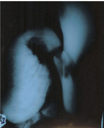
Figure 1: Upper GI series showing megaduodenum.
The aortomesentric angle was measured to be 12°. A provisional diagnosis of megaduodenum secondary to chronic SMA syndrome with an associated partial mid gut malrotation was made.
Gastric and duodenal decompression was performed pre-operatively.
At surgery, the duodenum was found to be grossly dilated extending upto the right iliac fossa. The caecum and ascending colon were in the normal anatomic position but had a mesocolon making them both mobile. A large left paraduodenal recess was found with internal herniation of the entire jejunum and part of the ileum (Figure 2). The ligament of Treitz could not be clearly defined. The recess was laid open and the kink at the duodenojejunal flexure was relieved. Following this, the position of the SMA
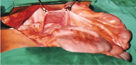
Figure 2: Intraoperative photograph showing a large paraduodenal recess.
Suggested continued obstruction of the duodenum necessitating a drainage procedure. A retrocolic, side to side, hand sewn, Roux-en-Y duodenojejunostomy was created at the most dependent part of the duodenum to allow adequate drainage of the area.
Post-operatively oral feeds were started on the fifth day. Post prandial vomiting was present. She settled down with prokinetics and was discharged on the tenth post-operative day. Thus, a final diagnosis of megaduodenum secondary to an internal hernia into a paraduodenal recess and chronic SMA syndrome was made.
Discussion
Megaduodenum is a rare entity in adults which may be either primary idiopathic or secondary. The secondary causes include Chagas disease, systemic sclerosis, duodenal stenosis, and visceral myopathy, connective tissue disorders such as Ehlers Danlos syndrome, strongyloidiasis [1] or superior mesenteric artery syndrome. Mid gut malrotation and congenital bands usually present in childhood. Adults usually present with abdominal pain, nausea, vomiting and complications of bacterial overgrowth: deconjugation of bile salts, malabsorption and polyneuropathy [2].
SMA syndrome usually occurs in older children and adolescents with a female preponderance. The SMA usually forms an angle of approximately 45° (range, 38- 56°) with the abdominal aorta, and the third part of the duodenum crosses caudal to the origin of the SMA, coursing between the SMA and aorta [3,4]. Any factor that sharply narrows the aortomesenteric angle to approximately 6-19° can cause entrapment and compression of the third part of the duodenum as it passes between the SMA and aorta, resulting in SMA syndrome [5]. In addition, the aortomesenteric distance in SMA syndrome is decreased. Alternatively, other causes implicated in SMA syndrome include high insertion of the duodenum at the ligament of Treitz, a low origin of the SMA, and compression of the duodenum due to peritoneal adhesions. The patient often presents with chronic upper abdominal symptoms such as epigastric pain, nausea, eructation, voluminous vomiting (bilious or partially digested food), postprandial discomfort, early satiety, and sometimes, sub-acute small- bowel obstruction [3]. The symptoms are typically relieved when the patient is in the left lateral decubitus, prone, or knee-to-chest position, and they are often aggravated when the patient is in the supine position [3].
Congenital internal hernias are unexpected and unusual lesions. They may be discovered as incidental findings at surgery or autopsy. They may remain silent or give rise to chronic digestive complaints or acute bowel obstruction. Internal hernias are paraduodenal in over fifty percent of cases, and three-fourths of these occur on the left side and they involve the paraduodenal fossa of Landzert as a result of a congenital defect in the fusion of the mesocolon and the mesentery of the duodenum with the peritoneum of the posterior abdominal wall [6]. Patients typically present acutely with small bowel obstruction. They often report a history of recurrent vague abdominal pain that has been present from months to years. Other complaints may include nausea, vomiting, and pain while standing. Symptoms may result from a partial small bowel obstruction with spontaneous reduction of the hernia. Symptoms are often postprandial, and pain may be relieved when the patient is supine.
Irrespective of the cause, the obstruction should be relieved. Many surgical techniques have been described for the repair of megaduodenum, including radical enterectomy, side-to-side duodenojejunostomy, duodenoplasty with feeding jejunostomy, duodenectomy with reimplantation of the ampulla of Vater and the roux-en-Y duodenojejunostomy. A tapering duodenoplasty can be performed to reduce the caliber of the duodenum [2].
In conclusion, megaduodenum is a rare anomaly in adults which poses a diagnostic and therapeutic challenge to the surgeon. Our patient had a megaduodenum secondary to a paraduodenal hernia compounded by chronic SMA syndrome, successfully treated with a Roux-en-Y duodenojejunostomy. The success of surgery relies on provision of adequate drainage. Once this is achieved, rapid alleviation of symptoms ensues along with a dramatic improvement in the quality of life.
8486
References
- Thompson BF, Fry LC, Wells CD, Olmos M, Lee DH, et al. (2004) The spectrum of GI strongyloidiasis: an endoscopic-pathologic study. GastrointestEndosc 59: 906-910.
- Nichol PF, Stoddard E, Lund DP, Starling JR (2004) Tapering duodenoplasty and Roux-en-Y duodenojejunostomy in the management of adult megaduodenum. Surgery 135: 222-224.
- Wilson-Storey D, MacKinlay GA (1986) The superior mesenteric artery syndrome. J R Coll Surg Edinb 31: 175-178.
- Hines JR, Gore RM, Ballantyne GH (1984) Superior mesenteric artery syndrome. Diagnostic criteria and therapeutic approaches. Am J Surg 148: 630-632.
- Shetty AK, Schmidt-Sommerfeld E, Haymon ML, Udall JN Jr (1999) Radiological case of the month. Superior mesenteric artery syndrome. Arch PediatrAdolesc Med 153: 303-304.
- Ovali GY, Orguc S, Unlu M, Pabuscu Y (2005) Transient left paraduodenal hernia. Comput Med Imaging Graph 29: 459-461.

