Marilia Arcadipane*, Luis Alberto Lage and Juliana Pereira
Universidade de Sao Paulo Faculdade de Medicina Hospital das Clinicas Instituto do Cancer São Paulo, São Paulo Brazil
- *Corresponding Author:
- Marilia Arcadipane
Universidade de Sao Paulo Faculdade
de Medicina Hospital das Clinicas
Instituto do Cancer São Paulo
São Paulo Brazil
Tel: +551145215505
E-mail: malcadipani@uol.com.br
Received Date: May 26, 2017 Accepted Date: June 21, 2017 Published Date: June 30, 2017
Citation: Arcadipane M, Lage LA, Pereira L (2017) Ascitic Fluid Cytology - Numerous Pleomorphic Lymphoid Cells in Primary Effusion Lymphoma. Ann Clin Lab Res. 5: 2.
Case Blog
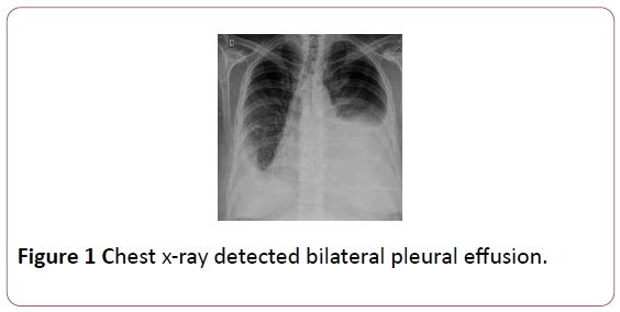
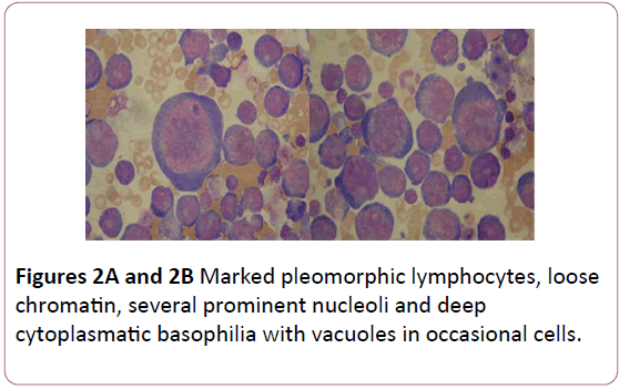
A 62-year-old man with a history of HIV/AIDS in regular antiretroviral therapy and CD4 count = 50 cells /mm3 and viral load=8340 copies/mm3, seek medical attention for fever, profuse night sweats and weight loss. Also, his complaints include dyspnea and progressive increase in abdominal volume. No lymphadenophathy was found. Blood smear showed normocytic anaemia (haemoglobin concentration 12 g/l), and chest x-ray detected bilateral pleural effusion (Figure 1). Abdominal CT scan showed massive ascites. A diagnostic and therapeutic paracentesis and thoracocentesis was performed, and peritoneal and pleural fluids analysis revealed exudates. Cytocentrifuge preparation of the patient’s ascitic fluid demonstrated massive cellular infiltrate composed of marked pleomorphic lymphocytes, loose chromatin, several prominent nucleoli and deep cytoplasmatic basophilia with vacuoles in occasional cells (Figures 2A and 2B). Flow cytometry analysis showed 32% of cells with hematopoietic phenotype activation kappa monoclonal CD45++, CD19-, CD20-, CD38++, CD56-, CD138-, CD3, cykappa+, and cylambdaexpression. Clinical and laboratory findings established a Primary effusion lymphoma (PEL) and due to poor performance status the patient was first treated with CVP but died of infectious complications before we could consider a more aggressive approach.
Figure 1: Chest x-ray detected bilateral pleural effusion.
Figure 2: Marked pleomorphic lymphocytes, loose chromatin, several prominent nucleoli and deep cytoplasmatic basophilia with vacuoles in occasional cells.
PEL is an aggressive large B cell neoplasm usually presenting with serous effusions and no detectable tumor masses. PEL etiopathogenesis is associated with herpes virus type 8 (HHV-8) infections and occurs in the setting of immunodeficiency, most commonly HIV infection in young or middle-aged homosexual males. Co-infection with Epstein-Barr virus (EBV) is frequent. Although most commom sites of involvement are the pleural, pericardium and peritoneum cavities, extracavitary involvement may occur, including the gastrointestinal tract, skin, lung or central nervous system. Up to 50% of cases have pre-existing Kaposi's sarcoma or may develop it subsequently to the diagnosis of lymphoma. Cytocentrifugation reveal large immunoblastic or plasmablastic cells, with prominent nucleoli and deeply basophilic cytoplasm. Occasionally vacuoles can be seen. Phenotypically there is CD45 expression, but pan-B antigens (CD19, CD20, and CD79a) are absent. Plasma cell differentiation markers and activation as CD38 and CD138 are often expressed as well as the latent membrane protein (LANA) associated the HHV8. This disease is AIDS defining and has an extremely poor prognosis, with median survival of 6 months, and poor response to chemotherapy with CHOP-like regimen.
19609


