Prashant Kumar Tripathi*
Vivekananda Polyclinic and Institute of Medical Sciences, Lucknow, Uttar Pradesh, India
*Corresponding Author:
Prashant Kumar Tripathi
Vivekananda Polyclinic and Institute of Medical Sciences, Vivekanandpuri Hydel Colony, Nirala Nagar, Lucknow, Uttar Pradesh-226007, India.
Tel: +918795582392
E-mail: tripathiprashant49@gmail.com
Received Date: October 15, 2017; Accepted Date: October 25, 2017; Published Date: October 31, 2017
Citation: Tripathi PK (2017) Cavernous Sinus Thrombosis Secondary to Retro-Orbital Abscess. J Neurol Neurosci Vol. 8 No: S4: i227.
Clinical Image
Cavernous sinus thrombosis is a serious condition that may be the result of any contigous septic focus. Its consequences can be fatal as well as lead to significant morbidity. I report a case of a young female who succumbed to unilateral cavernous sinus thrombosis secondary to ipsilateral retroorbital abscess. MRI brain and venogram were utilised to confirm the diagnosis.
A 25-years-old female came to the Neurology OPD with complaints of severe headache, high grade fever and multiple recurrent boils over left eye for 20 days. Gradually she developed proptosis of left eye with restriction of ocular movements in all directions. Her MRI of brain revealed mildly enlarged left cavernous sinus with interspersed non-enhancing foci likely due to thrombosis. MRI also revealed a small abscess posterior to the left eye. Her MRV Brain revealed impaired visualisation of left transverse sinus indicating its thrombosis (Figure 1).
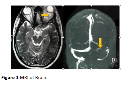
Figure 1: MRI of Brain.
The diagnosis of cavernous sinus thrombosis (CST) is based on clinical findings and confirmed by appropriate radiographic studies. MRI and venogram are more sensitive methods which have been proposed to reveal flow parameters. Findings may include a heterogenous signal from abnormal cavernous sinus and hyper intense signal of the thrombosed vascular sinuses, along with the deformity of the cavernous portion of internal carotid artery [1]. External ophthalmoplegia, defined as paralysis of extra-ocular muscles (in the case of CST, Secondary to dysfunction of third, fourth and sixth cranial nerves, rather than direct involvement of extra-ocular muscles), usually includes all the extra-ocular muscles [2].
20911
References
- Arian M, Kamali A, Abatabaeichehr M, Arashnia P (2016) Septic cavernous sinus thrombosis: A case report. Iran Red Crescent Med J 18: e34961.
- John RE, Mitchell TP, Niazi AF (2001) Septic thrombosis of the cavernous sinuses. Arch Intern Med 161: 2671-2676.

