Adeline Mei-YenYong*and Huma Jaffar
Department of Dermatology, National University Health System, Singapore
- *Corresponding Author:
- Adeline Mei-Yen Yong
Department of Dermatology, National University Health System
Singapore
Tel: 65 9623 6023
E-mail: adeline_meiyen@hotmail.com
Received date: March 26, 2016 Accepted date: March 28, 2016 Published date: March 31, 2016
Citation: Yong AMY, Jaffar H, et al. Cutaneous Sarcoidosis Treated as Seborrheic Dermatitis. Ann Clin Lab Res. 2016, 4:1.
Sarcoidosis is a multisystem disorder characterised by noncaseating granulomas in various organs and tissue. Common sites include the lymph nodes, lungs, spleen, liver, eyes, skin and joints. Cutaneous sarcoidosis is rare in Asia, and can occur in approximately 25 percent of patients, with vastly heterogeneous morphologic manifestations. Skin manifestations of sarcoidosis can be disfiguring, have prognostic importance, and may not be readily diagnosed even by dermatologists. We present a case of cutaneous sarcoidosis in Asia, initially treated as seborrheic dermatitis and responding to systemic therapy involving glucocorticoid and hydroxy chloroquine.
Introduction
Sarcoidosis is a multisystem disorder characterised by noncaseating granulomas in various organs and tissue. Common sites include the lymph nodes, lungs, spleen, liver, eyes, skin and joints. Cutaneous sarcoidosis is rare in Asia, and can occur in approximately 25 percent of patients, with vastly heterogeneous morphologic manifestations [1,2]. Skin manifestations of sarcoidosis can be disfiguring, have prognostic importance, and may not be readily diagnosed even by dermatologists [3]. We present a case of cutaneous sarcoidosis in Asia, initially treated as seborrheic dermatitis and responding to systemic therapy involving glucocorticoid and hydroxy chloroquine.
Image and Discussion
A 26-year -old Indian lady presented with a 5-month history of an itchy rash on the face. She was previously treated as seborrheic dermatitis with 1% Bethamethasone Valerate cream, Clobetasone and Tacrolimus ointment, however reported worsening of the rash. Review of systemic symptoms was unremarkable.
Physical examination revealed infiltrative erythematous papules and plaques over her eyelids and central face, including the nasolabial folds and perioral regions (Figures 1 and 2). The tip of the nose was spared. There were no annular lesions, nodules, crusting, pustules, telangiectasia or cervical lymphadenopathy. Rest of the cutaneous examination was unremarkable.
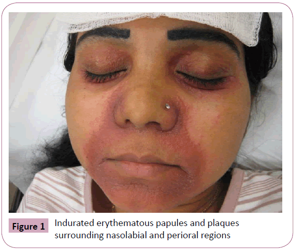
Figure 1: Indurated erythematous papules and plaques surrounding nasolabial and perioral regions
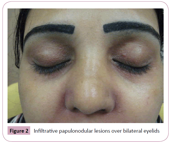
Figure 2: Infiltrative papulonodular lesions over bilateral eyelids
Skin biopsy over her left nasolabial fold revealed non-caseating granulomas with dermal aggregates of epithelioid histiocytes and lymphocytic multinucleated giant cells. Stains for acid fast bacilli (AFB), Fungus and Leprobacilli were negative, along with tissue cultures for AFB and fungal elements. There was no polarisable material detected. A rheumatology consult was sought to evaluate involvement of other organ systems. Eye examination, lung and renal function, chest radiograph and TB Quantiferon tests were normal.
Our patient was treated with daily Hydroxy chloroquine 200 mg and tapering doses of Prednisolone starting from 30 mg a day. There was 80% clearance of lesions at the end of 3 months without scarring.
Cutaneous sarcoidosis is notably rare in an Asian population, however it should be considered in patients with infiltrative lesions in a seborrheic distribution. In general, treatment of cutaneous sarcoidosis is reserved for patients with cosmetically disfiguring, symptomatic, ulcerating, or progressively worsening skin disease. Few randomized trials have compared the efficacy of treatment strategies [4-6]. In our patient, aggressive treatment with systemic therapy upon failing topical treatment may have caused the lesions to heal without scarring. Despite the low prevalence of sarcoidosis in Asia and an even smaller percentage of patients with cutaneous manifestations, attention must be spent on careful diagnosis and evaluation of systemic involvement. Further monitoring after treatment response is essential in view of possible relapse and subsequent systemic involvement [7,8].
8869
References
- Chong WS, Tan HH, Tan SH (2005) Cutaneous sarcoidosis in Asians: a report of 25 patients from Singapore. Clin Exp Dermatol 30: 120-124.
- Mangas C, Fernández-Figueras MT, FitéE, Fernández-Chico N, Sàbat M, et al. (2006) Clinical spectrum and histological analysis of 32 cases of specific cutaneous sarcoidosis. J Cutan Pathol 33: 772.
- Lodha S, Sanchez M, Prystowsky S (2009) Sarcoidosis of the skin: a review for the pulmonologist. Chest 136: 583.
- Baughman RP, Lower EE (2007) Evidence-based therapy for cutaneous sarcoidosis. Clin Dermatol 25: 334.
- Mosam A, Morar N (2004) Recalcitrant cutaneous sarcoidosis: an evidence-based sequential approach. J Dermatolog Treat 15: 353.
- Badgwell C, Rosen T (2007) Cutaneous sarcoidosis therapy updated. J Am Acad Dermatol 56: 69.
- Albertini JG, Tyler W, Miller OF (1997) Ulcerative sarcoidosis. Case report and review of the literature. 3rd. Arch Dermatol 133: 215.
- Mañá J, Marcoval J, Graells J, Salazar A, PeyríJ, et al. (1997) Cutaneous involvement in sarcoidosis. Relationship to systemic disease. Arch Dermatol 133: 882.


