Omer Engin1*, Oguzhan Sunamak2 , Ali Tosun3 , Mebrure Evnur Uyar4
1Surgery Department, Buca Seyfi Demirsoy State Hospital, Izmir, Turkey
2Surgery Department, Haydarpasa Numune Training and Research Hospital, Istanbul, Turkey
3Radiology Department, Sanliurfa Mehmet Akif Inan Training and Research Hospital, Sanliurfa, Turkey
4Emergency Department, Buca Seyfi Demirsoy State Hospital, Izmir, Turkey
- *Corresponding Author:
- Omer Engin, MD
General Surgeon Buca Seyfi Demirsoy State Hospital
Surgery Department
Izmir, Turkey
Tel: +902322505050
E-mail: omerengin@hotmail.com
The criminal events like stabbing are infrequent cases in surgical practice. The point of stabbing and the organs injured organs give the direction to the treatment. Thus, there are some diagnostic tools to determine which organs are injured. One of them is CT. CT with intravenous contrast helps us to see the trace of the stab wound and the degree of the injury. Also, CT images can be consulted to a radiologist via internet viewing systems, which is very useful for correct interpretation and removes the necessity of an on-duty radiologist in hospital
We used these properties of CT imaging in our case. The patient with the stabbing wound at right lumber area was taken with abdominal CT with IV contrast. The trace of stabbing and stabbingdependant hematoma at the level of the right paravertebral muscle was seen and it was not as deep as to enter into the abdominal cavity (Figures 1-3). Also, there was not any free air or fluid within the abdominal cavity. The patient was followed without operation and discharged 24 hours later without any complication. We want to emphasize that the use of CT and modern telecommunication facilities are important in early and correct diagnosis in such cases to avoid unnecessary operations [1-5].
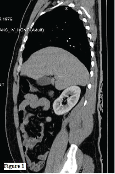
Figure 1:
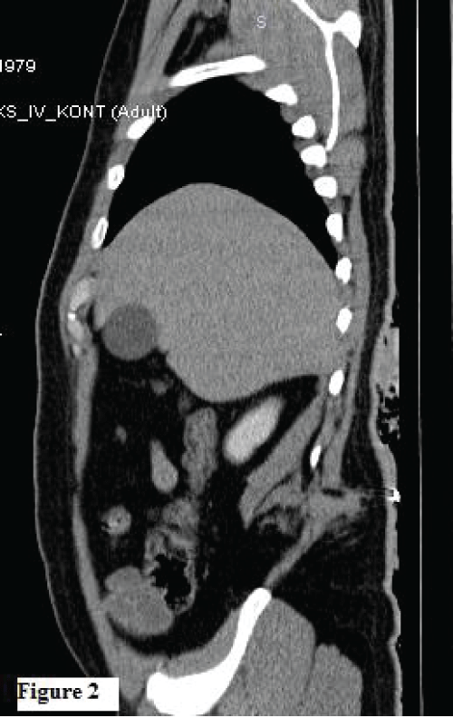
Figure 2:
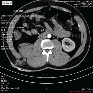
Figure 3:
6028
References
- Inaba K, Okoye OT, Rosenheck R, Melo N, Branco BC, et al. (2013) Prospective evaluation of the role of computed tomography in the assessment of abdominal stab wounds. JAMA Surg 148: 810-816.
- Bolliger SA, Ruder TD2, Ketterer T3, Gläser N4, Thali MJ5, et al. (2014) Comparison of stab wound probing versus radiological stab wound channel depiction with contrast medium. Forensic SciInt 234: 45-49.
- Bansal V, Reid CM, Fortlage D, Lee J, Kobayashi L, et al. (2014) Determining injuries from posterior and flank stab wounds using computed tomography tractography. Am Surg 80: 403-407.
- Lee GJ, Son G, Yu BC, Lee JN, Chung M (2014) Efficacy of computed tomography for abdominal stab wounds: a single institutional analysis. European Journal of Trauma and Emergency Surgery1-6.
- Engin O, Ipekci F, Calik B, Ucar AD, Tekin V (2012) Features of the cases injured by stab wounds. Journal of Universal Surgery1: 1-6.

