Introduction
|
| Due to susceptibility to fracture, restoration of maxillary anterior teeth after endodontic therapy can be a difficult task. Adequate restoration of anterior teeth is important for two reasons; 1) restoring the esthetic, and 2) function [1]. An endodontically treated tooth can be restored by using direct or indirect restorative techniques. However, in most cases restoration of these teeth will require post placement for retention of the core material and coronal reconstruction of the tooth [2]. Post retained final restorations are indicated when majority of tooth structure is lost due to decay [1]. |
| However, these posts have the disadvantages of being more expensive because they will need an intermediate laboratory phase for fabrication and more office visits. In addition, post placement increases the risk of the root fractures [3]. Also, they are not appropriate for esthetic zone because of their metallic reflection color [4]. Retention to the root canal is an important characteristic feature of the posts to protect the remaining structure [5]. Metallic cast posts have ragged surfaces that help them to have a strong retention, while glass-fiber posts have smooth surfaces. However, glass-fiber posts are more desirable because of their flexibility and resistance to vertical root fracture [1,3,6]. Airborne-particle abrasion of the surface of the post can improve the retention of glass-fiber post because it increases surface area, and enhances mechanical interlocking between the cement and roughened surface of the post [7]. Glass-fiber posts are made of 70% fiberglass, which is used to reinforce rigidity and resistance, and 30% of resin matrix that contains the fibers. These fibers are oriented parallel and in one direction to strengthen mechanical characteristics [6]. The modulus of elasticity of glassfiber posts is similar to that of dentin. So, the stress spreads equally along the tooth in comparison to more rigid posts [8]. Another characteristic feature of glass-fiber posts is that they can easily be removed from canals when the endodontically treated tooth has to be retreated [7,9,10]. Therefore, an important issue is to improve the retention of glass-fiber posts. Different studies have shown that physical or chemical treatments will improve the bond between cements and glass-fiber posts [11-13]. It is shown that physical treatment like changing the surface roughness by making rags on the surface of the posts is aggressive and could change the morphology of glass-fiber posts, and as a result it would interfere with the post fitness into the root canal [8]. In contrary, chemical treatments such as soaking the posts in special chemical solutions have advantages of being cheaper, less aggressive, and easier to apply. These solutions also clean the post surfaces [8]. There are lots of studies that have shown the effects of different factors on retention of glass-fiber posts into the canals [1,3,5,14-17]. |
| The aim of this in vitro study was to compare the retention of glass-fiber posts in endodontically treated teeth by using two different surface treatment methods in maxillary central incisors. |
Methodology
|
| In this in vitro study, forty-eight extracted non carious human maxillary central incisors with the average length of 22 to 22.5 millimeter (mm) were used [3,15,18]. These teeth had been extracted due to orthodontic therapy or periodontal problems after approval by the Ethics in Research Committee of Ahvaz Jundishapur University of Medical Sciences, Ahvaz, Iran. These teeth were preserved in physiological saline solution in 37?C before the experiment [18,19]. Before the teeth being send to the laboratory, Cavitron (30K TFI-10 6374106, Dentsply/Australia) was used to remove calculus and plaque from the surface of the teeth. Also, a Light Emission Diode (LED MID, Mident Industrial Co., Ltd/China) was used to ensure that these teeth have no caries lesions, superficial or deep cracks, or any crown and root fracture. |
| Based on the selection criteria mentioned above all forty-eight teeth deemed qualified to be part of the study. Access preparation was completed by using flat end cylinder diamond bur (K.G Sorernsen, Barueri/Brazil) with water spray just below the CEJ (Cemento-Enamel Junction) area to have a reference point for the length measurements in all teeth [16]. Step-back technique was used for canal preparation, K files (Mani/Japan) numbers 15 to 40 were used to clean the canals, and the K file number 40 was considered as the master apical file. Then, 1 through 4 numbers of the gates-Glidden (Lexicon, Dentsply/France) was used for shaping the canals. After shaping completed, the canals were irrigated with 1.5% sodium hypochlorite, and dried by paper points (Coltène Whaledent AG, Langenau/Germany) [18]. Obturation was completed by using lateral condensation technique with Gutta-Percha points (DiaDent Group International, ML 0.029, LOT# 010214, North Chungcheong/Korea), and root canal sealer,AH-26 silverfree (Dentsply DeTrey GmbH, LOT# 1310000224, Konstanz/Germany) [20]. |
| After obturation completed, the teeth were placed in physiological saline solution in 37?C [18]. The average root length of the central incisors used in this study was 12.5 mm. In the next step, post spaces were prepared to the depth of 8 mm leaving 4 mm of apical gutta-percha for an apical seal, by using a number 5 peeso- Reamer device (Mani/Japan) [2]. Then, the canals were cleaned by gentle water pressure, and dried by air pressure and a cotton wrapped file. Also, another cotton wrapped file imbrued with white ethanol was used to make sure that the whole canals are cleaned and dried. |
| After completion of the preparatory steps, the forty-eight teeth were divided into 3 groups of 16 teeth. In this study parallel intra radicular radiopaque glass-fiber posts with conical tips (Angelus, LOT#23229, Londrina/ Brazil) and different diameters were used. Three types of glass-fiber posts were prepared to be placed into the canals based on different diameters of teeth canals. The length of all posts was 20 mm. The small glass-fiber post had the diameter of 0.7 mm at the tip and 1.1 mm at the base. These numbers for the medium one was 0.9 and 1.3 mm, and for the large post was 1.1 and 1.5 mm. The first group of posts was the control group, and it had no superficial preparation (group A). The second group was prepared by sandblasting the surfaces of the posts under 60 Pascal pressure of Aluminum Oxide with 50 micrometer particles for 2 seconds (group B). The third group was placed in an ethanol solution for 1 minute (group C). |
| The next step was placing the glass-fiber posts into the canals with Panavia F2.0 cement (Kuraray Noritake Dental Inc. Tokyo/ Japan). The method for making the cement was based on the manufacturer instructions. Mixed cement was placed into the canals, and was gently spread with air pressure, and excess cement was removed via paper point. Next, a new mixture of the Panavia F 2.0 cement was made with the same ratio, and it was applied on the surfaces of the glass-fiber posts. These posts were placed at the center of the canals with a gentle finger pressure to hold the posts inside the canals for 6 minutes to allow completion of the cement auto-polymerization [18]. Then, cement was polymerized for 20 seconds with a polymerizing light placed 1 mm away from the surface of the tooth [18]. Teeth with cemented posts were then placed into the distilled water in 37?C for 24 hours [18]. This study did not use thermo cycling method for the teeth according to the studies of Porton et al. and Asakawa et al., because it has been shown that this method has no significant effect on the retention of posts into the canals [21,22]. Then, all the teeth were marked at 2 mm beneath the CEJ by using the surveying method, and they were mounted in a self-polymerizing acryl resin (Technovit, Heraeus Kulzer/Germany). Finally, a tensile force parallel to the long axis of the posts with a Universal Testing Machine (Zwick/ Roell, Type BDO-FB020TN, Ulm/Germany) at 0.5 mm per minute speed was applied to all of the posts [23]. Straining forces were continuously applied until post de-bonding from canals was clinically evident. The data of which included the maximum force needed to achieve de-bonding of the post was recorded and stored in Kilo Newton units (KN) by a computer device. |
|
Statistical Analysis
|
| The one-way analysis of variance (ANOVA) and Paired Sample Test were used for analyzing the results of this study, and comparison of the data. The significance level was set at p-Value 0.05. |
Results
|
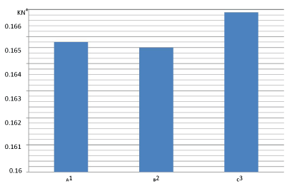
| Table 1 shows the maximum tensile strain that was used to move the posts out of the tooth canal. According to this table, the average of tensile strain for groups A and B was approximately the same (0.1648 and 0.1646). However, for group C, the average was a little higher than the two previous groups (0.1659). |
| According to Table 2, there is no significant difference among the three groups for minimum forces needed to dislodge the posts. This average was 0.132 KN for the group A, and 0.133 and 0.126 KN for groups B and C. Also, the maximum forces to move out the posts from the canals for groups A, B and C were 0.192, 0.196, and 0.2 KN. The standard deviations of tensile strain were small, and close to each other. |
| Table 3 states that there is no significant difference in p-value among the three groups (p-Value >0.05). |
| Figure 1 illustrates that differences of the average retention in these three groups are not remarkable. |
Discussion
|
| The primary goal of dentistry is to retain teeth as long as possible. When a large portion of the tooth is destructed due to dental decay or accidental fracture, preservation of remaining structure of the tooth is a major concern. Endodontics is a branch of dentistry, which offers a way to maintain teeth with irreversible pulpal damage for a longer time [2]. |
| Fiber posts are considered an alternative to metallic posts and cores in the restoration of endodontically treated teeth. The development of resin posts reinforced with glass or carbon fiber helped to minimize the difference between the modulus of elasticity of the restorative material and that of the tooth structure, to avoid root fracture [24]. The clinical success of glassfiber posts is most likely due to frictional retention between the post and tooth canal, instead of the effects of adhesive bonding [25]. The general agreement is that post retention is the major contributory factor in survival of the restorations [26,27]. |
| In this study, the sealer AH 26 was used for lateral condensation, and obturation of all forty-eight teeth. This sealer was selected because it contains no eugenol (studies have shown that eugenol inhibits the polymerization of luting agents with resin bases) [28,29]. This study had multiple outcomes. The Major outcome was that the average retention of glass-fiber posts in all three groups (A, B, and C) was close to 0.16 KN. This result is similar to the ones from the Gallo et al. study, who compared the average retention of fiber-composite posts and stainless steel posts, and demonstrated that the amount of retention for stainless steel posts with zinc phosphate cement was 0.43 KN; while this average for fiber-composite posts was 0.19 KN [17]. This negligible difference between two studies is maybe because of using different types of post and cement. |
| In addition, this study demonstrated that sandblasting the surface of the post before cementation does not improve the post retention. According to Sahafi et al., the retention of posts in anterior teeth is not always related to the surface preparation like sandblasting or silane application, and metal primer. He concluded that the type of the cement along with structure, and shape of the post has a major role in retention of posts into the canals [23]. Also, Soares et al. showed that airborne-particle abrasion has no effect on retention of posts into the canals, which confirms the result of this study [30]. However, Cheleux et al. has shown that sandblasting could enhance the bond strength of posts [31]. Another study, which is done by Radovic et al., avers that sandblasting might improve the microtensile strength of glass-fiber posts [32]. Moreover, Balbosh et al., Asakawa et al., and Choi et al. have shown that sandblasting with airborneparticle abrasion increases interlocking surfaces between posts and canals, which enhances the retention of posts considerably [7,22,33]. |
| This study also showed that placing the posts in ethanol solution before cementation does not increase the retention of posts into the canals in comparison with the control group. This result is consistent with the study of Cecchin et al. [34]. Although one study has implied that ethanol and dichloromethane improve bonding strength of the posts into the canals (Goncalves et al.) [8], other study, which is done by Asakawa et al. states that dichloromethane has no effect on micro tensile strength of glassfiber posts [22]. |
| The method of mechanical loading is a limitation of this study, which was parallel to the long axis of the posts. Furthermore, if restorations such as crown were used, the result of the study might be different. Also, it is not possible to achieve an exact simulation of the oral cavity environment. Factors such as type of the tooth (anterior or posterior), tooth structure remaining after decay removal, cement type, and esthetic considerations all play a major role in longevity and quality of post retention. Further studies on the retention of glass-fiber posts should be conducted to compare the retention of different post designs and varying surface treatments. |
Conclusion
|
| Based on these findings, and within the limitation of this study, no remarkable differences in retention of glass-fiber posts were observed between preparation with sandblasting and placing them in ethanol solution (p-Value >0.05). |
Acknowledgement
|
| We are very thankful to Dr. Nona Sanai for advice and assistance with editing. |
Tables at a glance
|
|
|
| |
Figures at a glance
|
 |
| Figure 1 |
|


