Armendáriz-Buil Ignacio1* and Gascón-Rubio María Cristina2
Department of Anesthesiology, Resuscitation and Pain Management, Hospital San Pedro, Logroño, Spain
Department of ENT, Hospital San Pedro, Logroño, Spain
*Corresponding Author:
Armendáriz-Buil Ignacio
Department of Anesthesiology, Resuscitation and Pain Management
Hospital San Pedro, Logroño
Spain
Tel: +34658643547
Fax: +34658643547
E-mail: iarmendariz@riojasalud.es
Received date: February 22, 2016; Accepted date: March 01, 2016; Published date: March 04, 2016
Citation: Ignacio AB, Cristina GRM. Foreign Body in Dorsolumbar Region Seen in Preoperative Radiological Study. Ann Clin Lab Res. 2016, 4:1.
Introduction
The image belongs to the preoperative study of a 65 yeas old patient scheduled to be operated on an ESS (Endoscopic Sinus Surgery). At the chest radiograph, one thread like metallic density of about 4.84 cm in length is identified in the subcutaneous tissue of the thoracolumbar region suggesting the presence of a foreign body, possibly a needle (Figures 1 and 2) image.
The patient was completely asymptomatic, the time of evolution was unknown and there were no previous image studies. Due to the absence of clinics, we decided a conservative approach and the intervention was carried out without any adverse event.
Case Report
We present the radiographic study of a 65 years old woman diagnosed of sinonasal polyposis grade III scheduled for ESS. The patient reported no drug allergies, no previous surgeries and her only medical illness were hypertension.
Following the protocol of our hospital, a complete pre-operative testing was completed before going to the anaesthesia consultation. This study includes analytics, electrocardiogram and chest radiography.
The chest radiography revealed an elongate foreign body with metallic density: possibly a needle in the subcutaneous tissue (Figures 1 and 2). At the anaesthesia consultation, the finding was communicated to the patient and she was questioned about its origin. The patient denied knowing the possible origin: no previous interventions, no acupuncture practitioner and the only possible source could be her love of sewing, but she did not remember ever having a needle stuck.
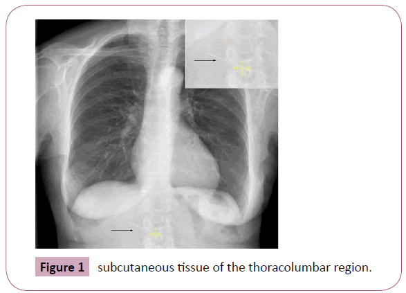
Figure 1: subcutaneous tissue of the thoracolumbar region.
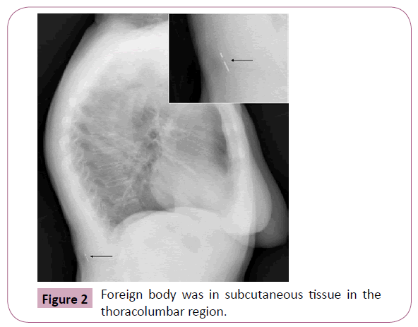
Figure 2: Foreign body was in subcutaneous tissue in the thoracolumbar region.
Due to the absence of symptoms and no implications for the development of the surgery, it was decided to be conservative. The intervention was performed under general anaesthesia without any notable event. The patient was discharged within 24 hours without incidents.
We advised the patient to communicate the presence of the foreign body in case to need health care.
Discussion
Aspired or ingested foreign bodies are the most often found at the chest x-ray, especially in children [1]. Foreign body ingestion is common problem that often requires little intervention [2].
Nevertheless, foreign body aspiration is often a serious medical condition demanding timely recognition and prompt action [3]. In the case presented, the foreign body was in subcutaneous tissue in the thoracolumbar region (Figure 2). This location is usually associated with acupuncture needles, placed permanently deliberately or inadvertently forgotten. They are usually inserted in paramedian location of the lumbar region [4]. Therefore, if inadvertently be forgotten, the paramedian location is the most common. However, in the case described, the needle was in medial location. On thoracolumbar region. Furthermore, acupuncture needles are typically about 1 mm in diameter and 10-15 mm in length [5], while the needle of our case was longer (48 mm). The size and location of the needle fits more with an accidental puncture of a sewing needle.
We decided a conservative approach since in most cases these needles may remain in situ forever without problems [4]. Although these needles occasionally migrate, especially in patients without much body fat, they usually do not result in complications [6]. Likewise, the prevalence of complications related to permanently retained needles remains unknown. There remains a paucity of literature describing such complications. When these retained needles appear during medical imaging, they are largely regarded as a medical curiosity. However, most of times, they have no clinical implications.
8754
References
- Passali D, Lauriello M, Bellussi L (2010) Foreign body inhalation in children: an update. ActaOtorhinolaryngolItal 30:27-32.
- Baharloo F, Veyckemans F, Francis C (1999) Tracheobronchial foreign bodies: presentation and management in children and adults. Chest 115:1357-1362.
- Qureshi A, Behzadi A (2008) Foreign-body aspiration in an adult. Can J Surg 51: 69-70.
- Galbraith P, Richardson M (2006) Permanently Retained Acupuncture Needles: Radiographic Findings and Case Report. Radiology Case Reports 1:120-122.
- Galuten A, Austin JH (1988) Permanent subcutaneous acupuncture needles: radiographic manifestations. Can AssocRadiol J 39:54-56.
- Gerard PS, Wilck E, Schiano T (1993) Imaging implications in the evaluation of permanent needle acupuncture. Clin Imaging 17:36-40.


