Flyer

Journal of Universal Surgery

  • ISSN: 2254-6758
  • Journal h-index: 6
  • Journal CiteScore: 0.94
  • Journal Impact Factor: 0.82
  • Average acceptance to publication time (5-7 days)
  • Average article processing time (30-45 days) Less than 5 volumes 30 days
    8 - 9 volumes 40 days
    10 and more volumes 45 days
Awards Nomination 20+ Million Readerbase
Indexed In
  • Genamics JournalSeek
  • Directory of Research Journal Indexing (DRJI)
  • OCLC- WorldCat
  • Euro Pub
  • Google Scholar
  • J-Gate
  • SHERPA ROMEO
  • International Committee of Medical Journal Editors (ICMJE)
  • Zenodo
Share This Page

Case Report - (2015) Volume 3, Issue 2

Hereditary Hemorrhagic Telangiectasia: Presenting with Epistaxis

Sandeep Kumar Kar1*, Manasij Mitra2,Tanmoy Ganguly1,Manabendra Sarkar3,Chaitali Sen Dasgupta1, Anupam Goswami1
  1. Department of Cardiac Anaesthesiology, Institute of Postgraduate Medical Education & Research, Kolkata, India
  2. Department of Anesthesiology, MGM Medical College Krisanjanj, India
  3. Department of Anaesthesiology, N.R.S Medical College Kolkata, India
Corresponding Author: Dr. Sandeep Kumar Kar, Assistant Professor, Dr. Sandeep Kumar Kar, Assistant Professor, Department of Cardiac Anaesthesiology Institute of Postgraduate Medical Education& Research, Kolkata, India,E-mail: sndpkar@yahoo.co.in
Related article at Pubmed, Scholar Google
Visit for more related articles at Journal of Universal Surgery

Abstract

Osler-Weber-Rendu disease (OWRD) or Hereditary Hemorrhagic Telangiectasia (HHT) is a rare autosomal dominant disorder that causes muco-cutanesous and visceral vascular dysplasia and results in increased tendency for bleeding [1-4]. Patients with HHT may present with variety of symptoms and management differs accordingly. Epistaxis is the most common symptom of HHT and mucocutaneous telangiectasia the most common sign [5]. Here we describe the anesthesia management of a patient presenting with epistaxis in emergency suffering from this syndrome.


Introduction

Osler-Weber-Rendu disease (OWRD) or Hereditary Hemorrhagic Telangiectasia (HHT) is a rare autosomal dominant disorder that causes muco-cutanesous and visceral vascular dysplasia and results in increased tendency for bleeding [1-4]. Patients with HHT may present with variety of symptoms and management differs accordingly. Epistaxis is the most common symptom of HHT and mucocutaneous telangiectasia the most common sign [5]. Here we describe the anesthesia management of a patient presenting with epistaxis in emergency suffering from this syndrome.

Case Report

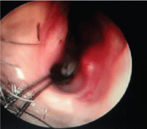
A 50 year old male patient of Indian origin came to the emergency with severe epistaxis. Past history and records revealed recurrent epistaxis with definitive positive family history and telangiectasia over nasal mucosa and paranasal sinuses. He had been diagnosed previously as HHT due to presence of vascular malformation in the nose and paranasal sinuses by endoscopy and stomach by upper GI endoscopy. After admission his blood pressure was 90/60 mm Hg, pulse 124/minute, room air SaO2 96%. Investigation revealed Hb 6.5 gm/dl, PCV 16 gm/dl. Chest auscultation revealed no crepitation and bilateral vesicular breath sound was present. He was stabilised with 500 ml hydroxyl-ethyl starch till blood products were made available, followed by 3 unit packed cell and 4 unit FFP transfusion. Patient received tranexamic acid 1 gm slow I.V infusion to control the bleeding, glycopyrrolate to reduce secretions, pantoprazole as antiulcer prophylaxis and Ondansetron to reduce reflux. Along with resuscitation, surgical exploration with control of bleeding under general anesthesia was planned. As blood in stomach was a possibility and patient’s fasting status was unknown, rapid sequence induction was carried out with thiopentone 5 mg/kg and succinylcholine 2 mg/ kg and trachea was intubated with a 8.5 mm I.D. cuffed portex endotracheal tube. A urinary catheter was inserted and orogastric tube aspiration was done. Maintenance of anesthesia was done with sevoflurane, nitrous oxide and oxygen, and atracurium as a muscle relaxant. Several telangectasias were found over the anterior part of the nasal septum which was thoroughly cauterized with diathermy under endoscopic guidance. This was followed by septo-dermoplasty to stop the bleeding. Intraoperatively tranexamic acid was given (1 gm) along with local packing with lignocaine and adrenaline mixture. Intraoperatively controlled hypotension was induced with sevoflurane and propofol boluses to maintain systolic blood pressure between 90 to 100 mmHg. Patient was reversed by using standard reversal techniques with neostigmine and glycopyrollate. Postoperative period was uneventful (Figure 1).

Discussion

HHT is a rare systemic fibro vascular dysplasia [6] with incidence varying from 1 in 5,000 to 10,000 [7] to 1 to 2 in 1,00,000 [6]. Sutton [8] in 1864 first described this syndrome in a man with a vascular malformation and recurrent epistaxis. In 1896 Rendu [9] first noted the association between hereditary epistaxis and telangiectasia in a 52 years old man. Osler [10] in 1901 and Weber [11] in 1907 further elaborated the association between hemorrhagic lesions in skin and mucous membranes and its familial inheritance. Although the disease is popularly known as Osler-Weber-Rendu syndrome, the name ‘hereditary hemorrhagic telangiectasia’ suggested by Hanes [12] in 1909, recognizes the characteristics that define the disease.
HHT is manifested by mucocutaneous telangiectases and arteriovenous malformations (AVMs) in different parts of body. Lesions can affect the nasopharynx, central nervous system (CNS), lung, liver, and spleen, as well as the urinary tract, gastrointestinal (GI) tract, conjunctiva, trunk, arms, and fingers [2,13]. Impaired signalling of transforming growth factor-ß/bone morphogenesis protein (TGF-β/BMP) [14-17] as well as vascular endothelial growth factor (VEGF) [18,19] has been attributed as the primary cause of HHT. The gene mutations found to be responsible are as mentioned in Table 1.
“definite” if 3 or more criteria are present, “possible or suspected” if 2 criteria are present, and “unlikely” if 0 or 1 criterion is present.
The diagnosis of HHT is made clinically on the basis of the Curaçao criteria [3], established in June 1999 by the Scientific Advisory Board of the HHT Foundation International, Inc. The 4 clinical diagnostic criteria are as follows:
HHT Foundation International - Guidelines Working Group [20] has recommended diagnosis of HHT using the Curaçao Criteria (Table 2) or by identification of a causative mutation.
Histopathology of HHT lesions show many layers of smooth muscle cells without elastic fibers and very frequently arterioles directly communicating with smooth muscle cells. As a result telangiectases are very sensitive to slight trauma and friction. HHT may present in children as bleeding but usual age of presentation in adulthood [4]. Male and females are equally affected [21]. Classic triad of presentation include telangiectases of the skin and mucous membranes, epistaxis, and a positive family history. Epistaxis may be present in upto 95% cases [4,22] whereas skin lesions account for 75-90% of presentations [22,23]. Skin telangiectasias rarely cause bleeding [4]. Gastrointestinal telangiectasia may occur in 10-33% patients [24] most commonly in the stomach and upper duodenum [24]. Significant bleeding from gastrointestinal tract may occur in 25% patients older than 60 years and may increase with age [25]. Pulmonary involvement in the form of arteriovenous malformations (AVMs) may be present in 75% HHT1 and 44% HHT2 patients [26]. Patients with pulmonary involvement are at high risk of developing cerebral thrombotic and embolic events including stroke, brain abscess, or transient ischemic attacks due to right-to-left shunting [14,24]. Cerebral AVMs may be present 15-20% HHT1 and 1-2% HHT2 patients [26-30], and may present with seizure, headache or intracranial haemorrhages [4,31]. Hepatic AVMs may be present upto 74% cases [32] but usually asymptomatic [4]. Management strategies for AVMs associated with HHT may differ with location and presentation and depicted in Table 3.
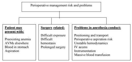
Patients with HHT presenting with continuous bleeding pose a serious problem to the anaesthesiologists. Pre-existing anemia due to recurrent bleeding is common and sudden decompensation may lead to heart failure. Uncontrolled bleeding may occur from skin lesions during patient positioning and transport. Epistaxis may lead to aspiration of blood into trachea causing pulmonary edema. Intravenous access may be difficult. Sudden change in blood pressure may cause bleeding from AVMs anywhere in the body, most serious of which is from cerebral AVM. Gastric distension may occur from ingested blood and may cause reflux and aspiration during induction. Any instrumentation including laryngoscopy and intubation, nasogastric tube insertion, urinary catheterisation should be carried out with utmost caution as bleeding may occur from undetected lesions (Figure 2).
In stable patients, posted for elective surgery, preoperative optimization with oral or parenteral iron and if necessary erythropoiesis-stimulating agent [33] should be considered. Preoperatively angiogenesis inhibitor or hormone therapy should be considered in selected patients to reduce perioperative bleeding. Careful history and physical examination may indicate any systemic involvement and standard radiological imaging with angiography may be performed to search for hemangiomas in brain, lung, gastrointestinal tract, nose and paranasal sinuses. In unstable patient presenting with severe bleeding focus should be directed to simultaneous resuscitation and hemostasis. Blood transfusion forms the mainstay of volume resuscitation in severely volume depleted patient. Epistaxis should be controlled with tight nasal packing immediately followed by cauterisation of bleeding vessels and dermoplasty if required. Since bleeding does not result from a defect in coagulation cascade, but from the malformed vascular structures, platelet or plasma transfusions are of no use and reserved only to supplement the loss. Antifibrinolytics including tranexamic acid [34,35] and aminocaproic acid [36] have been used with success to control epistaxis. In addition to antifibrinolytic effects, tranexamic acid also stimulates the expression of ALK-1 and endoglin, as well as the activity of the ALK-1/endoglin pathway [37-45]. Intraoperatively controlled hypotension should be used to reduce bleeding.

Conclusion

Patients with Osler-Weber-Rendu disease (OWRD) or Hereditary Hemorrhagic Telangiectasia (HHT) may present with uncontrolled bleeding. Resuscitation alongwith hemostasis forms the cornerstone of treatment. As the bleeding occurs from malformed vessels standard coagulation tests will reveal no abnormality. Management include blood transfusion, antifibrinolytics and surgical hemostasis. Anesthesia strategy should include rapid sequence induction and controlled hypotension.

Tables at a glance

Table icon Table icon Table icon
Table 1 Table 2 Table 3

Figures at a glance

Figure 1 Figure 2
Figure 1 Figure 2
6034

References

  1. Peery WH (1987) Clinical spectrum of hereditary hemorrhagic telangiectasia (Osler-Weber-Rendu disease). See comment in PubMed Commons below Am J Med 82: 989-997.
  2. Guttmacher AE, Marchuk DA, White RI Jr (1995) Hereditary hemorrhagic telangiectasia. See comment in PubMed Commons below N Engl J Med 333: 918-924.
  3. Shovlin CL, Guttmacher AE, Buscarini E, Faughnan ME, Hyland RH, et al. (2000) Diagnostic criteria for hereditary hemorrhagic telangiectasia (Rendu-Osler-Weber syndrome). See comment in PubMed Commons below Am J Med Genet 91: 66-67.
  4. Sharathkumar AA, Shapiro A (2008) Hereditary haemorrhagic telangiectasia. See comment in PubMed Commons below Haemophilia 14: 1269-1280.
  5. Porteous ME, Burn J, Proctor SJ (1992) Hereditary haemorrhagic telangiectasia: a clinical analysis. See comment in PubMed Commons below J Med Genet 29: 527-530.
  6. Goulart AP, Moro ET, Guasti VM, Colares RF (2009) Anesthetic management of a patient with hereditary hemorrhagic telangiectasia (Rendu-Osler-Weber syndrome). Case report. See comment in PubMed Commons below Rev Bras Anestesiol 59: 74-78.
  7. Kjeldsen AD, Vase P, Green A (1999) Hereditary haemorrhagic telangiectasia: a population-based study of prevalence and mortality in Danish patients. See comment in PubMed Commons below J Intern Med 245: 31-39.
  8. Sutton H (1864) Epistaxis as an indication of impaired nutrition, and of degeneration of the vascular system. Med Mirror1: 769-781.
  9. Rendu H (1896) Epistaxis repetees chez unsujetporteur de petitsangiomescutanes et muquez. Gazette des HopitauxCivilsetMilitaires135: 1322-1323.
  10. Osler W (1901)On a family form of recurring epistaxis, associated with multiple telangiectases of the skin and mucous membranes. Bull Johns Hopkins Hosp 12: 333-337.
  11. Weber F (1907)Multiple hereditary developmental angiomata (telangiectases) of the skin and mucous membranes associated with recurring haemorrhages. Lancet 2: 160–162.
  12. Hanes F (109) Multiple hereditary telangiectases causing hemorrhage (hereditary hemorrhagic telangiectasia). Bull Johns Hopkins Hosp 20: 63-73.
  13. Nanda S, Bhatt SP (2009) Hereditary hemorrhagic telangiectasia: epistaxis and hemoptysis. See comment in PubMed Commons below CMAJ 180: 838.
  14. Shovlin CL, Letarte M (1999)Hereditaryhaemorrhagic telangiectasia and pulmonary arteriovenous malformations: issues in clinical management and review of pathogenic mechanisms. Thorax54: 714-729.
  15. tenDijke P, Hill CS (2004) New insights into TGF-beta-Smadsignalling. See comment in PubMed Commons below Trends BiochemSci 29: 265-273.
  16. Wrana JL, Attisano L, Wieser R, Ventura F, Massagué J (1994) Mechanism of activation of the TGF-beta receptor. See comment in PubMed Commons below Nature 370: 341-347.
  17. Schmierer B, Hill CS (2007) TGFbeta-SMAD signal transduction: molecular specificity and functional flexibility. See comment in PubMed Commons below Nat Rev Mol Cell Biol 8: 970-982.
  18. Cirulli A, Liso A, D'Ovidio F, Mestice A, Pasculli G, et al. (2003) Vascular endothelial growth factor serum levels are elevated in patients with hereditary hemorrhagic telangiectasia. See comment in PubMed Commons below ActaHaematol 110: 29-32.
  19. Xu B, Wu YQ, Huey M, Arthur HM, Marchuk DA, et al. (2004) Vascular endothelial growth factor induces abnormal microvasculature in the endoglin heterozygous mouse brain. J Cereb Blood Flow Metab 24: 237-244.
  20. McAllister KA, Grogg KM, Johnson DW, Gallione CJ, Baldwin MA, et al. (1994) Endoglin, a TGF-beta binding protein of endothelial cells, is the gene for hereditary haemorrhagic telangiectasia type 1. See comment in PubMed Commons below Nat Genet 8: 345-351.
  21. Abdalla SA, Letarte M (2006) Hereditaryhaemorrhagic telangiectasia: current views on genetics and mechanisms of disease. See comment in PubMed Commons below J Med Genet 43: 97-110.
  22. McDonald MT, Papenberg KA, Ghosh S, Glatfelter AA, Biesecker BB, et al. (1994) A disease locus for hereditary haemorrhagic telangiectasia maps to chromosome 9q33-34. See comment in PubMed Commons below Nat Genet 6: 197-204.
  23. Shovlin CL, Hughes JM, Tuddenham EG, Temperley I, Perembelon YF, et al. (1994) A gene for hereditary haemorrhagic telangiectasia maps to chromosome 9q3. See comment in PubMed Commons below Nat Genet 6: 205-209.
  24. Johnson DW, Berg JN, Baldwin MA, Gallione CJ, Marondel I, et al. (1996) Mutations in the activin receptor-like kinase 1 gene in hereditary haemorrhagic telangiectasia type 2. See comment in PubMed Commons below Nat Genet 13: 189-195.
  25. Johnson DW, Berg JN, Gallione CJ, McAllister KA, Warner JP, et al. (1995) A second locus for hereditary hemorrhagic telangiectasia maps to chromosome 12. See comment in PubMed Commons below Genome Res 5: 21-28.
  26. Cole SG, Begbie ME, Wallace GM, Shovlin CL (2005) A new locus for hereditary haemorrhagic telangiectasia (HHT3) maps to chromosome 5. See comment in PubMed Commons below J Med Genet 42: 577-582.
  27. Bayrak-Toydemir P, McDonald J, Akarsu N, Toydemir RM, Calderon F, et al. (2006) A fourth locus for hereditary hemorrhagic telangiectasia maps to chromosome 7. See comment in PubMed Commons below Am J Med Genet A 140: 2155-2162.
  28. Gallione CJ, Repetto GM, Legius E, Rustgi AK, Schelley SL, et al. (2004)A combined syndrome of juvenile polyposis and hereditary haemorrhagic telangiectasia associated with mutations in MADH4 (SMAD4). Lancet363: 852-859.
  29. Gallione C, Aylsworth AS, Beis J, Berk T, Bernhardt B, et al. (2010) Overlapping spectra of SMAD4 mutations in juvenile polyposis (JP) and JP-HHT syndrome. See comment in PubMed Commons below Am J Med Genet A 152A: 333-339.
  30. Iyer NK, Burke CA, Leach BH, Parambil JG (2010) SMAD4 mutation and the combined syndrome of juvenile polyposis syndrome and hereditary haemorrhagic telangiectasia. See comment in PubMed Commons below Thorax 65: 745-746.
  31. Harrison RE, Flanagan JA, Sankelo M, Abdalla SA, Rowell J, et al. (2003) Molecular and functional analysis identifies ALK-1 as the predominant cause of pulmonary hypertension related to hereditary haemorrhagic telangiectasia. J Med Genet 40: 865-871.
  32. Trembath RC, Thomson JR, Machado RD, Morgan NV, Atkinson C, et al. (2001) Clinical and molecular genetic features of pulmonary hypertension in patients with hereditary hemorrhagic telangiectasia. N Engl J Med345: 325-334.
  33. Faughnan ME,Palda VA, Garcia-Tsao G, Geisthoff UW, McDonald J, et al. (2011) HHT Foundation International - Guidelines Working Group. International guidelines for the diagnosis and management of hereditary haemorrhagic telangiectasia. J Med Genet 48: 73-87.
  34. Schoen FJ, Cotran RS, Vinay K, Collins T (1994) Robbins Pathologic Basis of Disease. 5th. WB Saunders.
  35. Plauchu H, de Chadarévian JP, Bideau A, Robert JM (1989) Age-related clinical profile of hereditary hemorrhagic telangiectasia in an epidemiologically recruited population. See comment in PubMed Commons below Am J Med Genet 32: 291-297.
  36. Berg J, Porteous M, Reinhardt D, Gallione C, Holloway S, et al. (2003)Hereditaryhaemorrhagic telangiectasia: a questionnaire based study to delineate the different phenotypes caused by endoglin and ALK1 mutations. J Med Genet 40: 585-590.
  37. Ingrosso M, Sabbà C, Pisani A, Principi M, Gallitelli M, et al. (2004) Evidence of small-bowel involvement in hereditary hemorrhagic telangiectasia: a capsule-endoscopic study. See comment in PubMed Commons below Endoscopy 36: 1074-1079.
  38. Kjeldsen AD, Kjeldsen J (2000) Gastrointestinal bleeding in patients with hereditary hemorrhagic telangiectasia. See comment in PubMed Commons below Am J Gastroenterol 95: 415-418.
  39. Sabbà C, Pasculli G, Lenato GM, Suppressa P, Lastella P, et al. (2007) Hereditary hemorrhagic telangiectasia: clinical features in ENG and ALK1 mutation carriers. See comment in PubMed Commons below J ThrombHaemost 5: 1149-1157.
  40. Vase P, Holm M, Arendrup H (1985) Pulmonaryarteriovenous fistulas in hereditary hemorrhagic telangiectasia. See comment in PubMed Commons below Acta Med Scand 218: 105-109.
  41. Haitjema T, Disch F, Overtoom TT, Westermann CJ, Lammers JW (1995) Screening family members of patients with hereditary hemorrhagic telangiectasia. See comment in PubMed Commons below Am J Med 99: 519-524.
  42. Fulbright RK, Chaloupka JC, Putman CM, Sze GK, Merriam MM, et al. (1998) MR of hereditary hemorrhagic telangiectasia: prevalence and spectrum of cerebrovascular malformations. See comment in PubMed Commons below AJNR Am J Neuroradiol 19: 477-484.
  43. Bossler AD, Richards J, George C, Godmilow L, Ganguly A (2006) Novel mutations in ENG and ACVRL1 identified in a series of 200 individuals undergoing clinical genetic testing for hereditary hemorrhagic telangiectasia (HHT): correlation of genotype with phenotype. Hum Mutat27: 667-675.
  44. Matsubara S, Mandzia JL, terBrugge K, Willinsky RA, Faughnan ME (2000) Angiographic and clinical characteristics of patients with cerebral arteriovenous malformations associated with hereditary hemorrhagic telangiectasia. AJNR Am J Neuroradiol 21: 1016-1020.
  45. Memeo M, Stabile Ianora AA, Scardapane A, Buonamico P, Sabbà C, et al. (2004) Hepatic involvement in hereditary hemorrhagic telangiectasia: CT findings. See comment in PubMed Commons below Abdom Imaging 29: 211-220.
  46. Boyer H, Fernandes P, Duran O, Hunter D, Goding G (2011) Office-based sclerotherapy for recurrent epistaxis due to hereditary hemorrhagic telangiectasia: a pilot study. Int Forum Allergy Rhinol1: 319-323.
  47. Mortuaire G, Boute O, Hatron PY, Chevalier D (2013) Pilot study of submucosal radiofrequency for epistaxis in hereditary hemorrhagic telangiectasia. See comment in PubMed Commons below Rhinology 51: 355-360.
  48. Dheyauldeen S, OstertunGeirdal A, Osnes T, Vartdal LS, Dollner R (2012) Bevacizumab in hereditary hemorrhagic telangiectasia-associated epistaxis: effectiveness of an injection protocol based on the vascular anatomy of the nose. Laryngoscope122: 1210-1214.
  49. Pau H, Carney AS, Murty GE (2001)Hereditaryhaemorrhagic telangiectasia (Osler-Weber-Rendu syndrome): otorhinolaryngological manifestations. ClinOtolaryngol Allied Sci, 2001; 26: 93-98.
  50. Strother CM, Newton TH (1976) Percutaneous embolization to control epistaxis in Rendu-Osler-Weber disease. See comment in PubMed Commons below Arch Otolaryngol 102: 58-60.
  51. Harries PG, Brockbank MJ, Shakespeare PG, Carruth JA (1997) Treatment of hereditary haemorrhagic telangiectasia by the pulsed dye laser. See comment in PubMed Commons below J LaryngolOtol 111: 1038-1041.
  52. Werner A, Bäumler W, Zietz S, Kühnel T, Hohenleutner U, Landthaler M (2008) Hereditary haemorrhagic telangiectasia treated by pulsed neodymium:yttrium-aluminium-garnet (Nd:YAG) laser (1,064 nm). Lasers Med Sci23: 385-391.
  53. vanCutsem E, Rutgeerts P, Vantrappen G (1990) Treatment of bleeding gastrointestinal vascular malformations with oestrogen-progesterone. See comment in PubMed Commons below Lancet 335: 953-955.
  54. Massoud OI, Youssef WI, Mullen KD (2004) Resolution of hereditary hemorrhagic telangiectasia and anemia with prolonged alpha-interferon therapy for chronic hepatitis C. See comment in PubMed Commons below J ClinGastroenterol 38: 377-379.
  55. Shovlin CL, Winstock AR, Peters AM, Jackson JE, Hughes JM (1995) Medical complications of pregnancy in hereditary haemorrhagic telangiectasia. See comment in PubMed Commons below QJM 88: 879-887.
  56. White RI Jr, Pollak JS, Wirth JA (1996)Pulmonaryarteriovenous malformations: diagnosis and transcatheterembolotherapy. J VascIntervRadiol7: 787-804.
  57. Hadjipanayis CG, Levy EI, Niranjan A, Firlik AD, Kondziolka D, et al. (2001) Stereotactic radiosurgery for motor cortex region arteriovenous malformations. See comment in PubMed Commons below Neurosurgery 48: 70-76.
  58. Levy EI, Niranjan A, Thompson TP, Scarrow AM, Kondziolka D, et al. (2000) Radiosurgery for childhood intracranial arteriovenous malformations. See comment in PubMed Commons below Neurosurgery 47: 834-841.
  59. Dupuis-Girod S, ChesnaisAL, Ginon I, Dumortier J, Saurin JC, et al. (2010) Long-term outcome of patients with hereditary hemorrhagic telangiectasia and severe hepatic involvement after orthotopic liver transplantation: a single-center study. Liver Transpl 16: 340-347.
  60. Hillert C, Broering DC, Gundlach M, Knoefel WT, Izbicki JR, et al. (2001) Hepatic involvement in hereditary hemorrhagic telangiectasia: an unusual indication for liver transplantation. Liver Transpl 7: 266-268
  61. Lerut J, Orlando G, Adam R, Sabbà C, Pfitzmann R, et al. (2006) Liver transplantation for hereditary hemorrhagic telangiectasia: Report of the European liver transplant registry. See comment in PubMed Commons below Ann Surg 244: 854-862.
  62. Mitchell A, Adams LA, MacQuillan G, Tibballs J, vandenDriesen R, et al. (2008) Bevacizumab reverses need for liver transplantation in hereditary hemorrhagic telangiectasia. See comment in PubMed Commons below Liver Transpl 14: 210-213.
  63. Cherif H, Karlsson T (2014) Combination treatment with an erythropoiesis-stimulating agent and intravenous iron alleviates anaemia in patients with hereditary haemorrhagic telangiectasia. Ups J Med Sci 119: 350-353.
  64. Gaillard S, Dupuis-Girod S, Boutitie F, Rivière S, Morinière S, et al. (2014)Tranexamic acid for epistaxis in hereditary hemorrhagic telangiectasia patients: a European cross-over controlled trial in a rare disease. J ThrombHaemost 12: 1494-1502.
  65. Morales-Angulo C, Pérez del Molino A, ZarrabeitiaR, Fernández A, Sanz-Rodríguez F, Botella LM (2007) [Treatment of epistaxes in hereditary haemorrhagic telangiectasia (Rendu-Osler-Weber disease) with tranexamic acid]. [Article in Spanish] ActaOtorrinolaringolEsp58: 129-1232.
  66. Korzenik JR, Topazian MD, White R (1994) Treatment of bleeding in hereditary hemorrhagic telangiectasia with aminocaproic acid. See comment in PubMed Commons below N Engl J Med 331: 1236.
  67. Fernandez-L A, Garrido-Martin EM, Sanz-Rodriguez F, Ramirez JR, Morales-Angulo C, et al. (2007) Therapeutic action of tranexamic acid in hereditary haemorrhagic telangiectasia (HHT): regulation of ALK-1/endoglin pathway in endothelial cells. ThrombHaemost 97: 254-262.