Fuat , Muharrem Karaoølan1 and Mehmet Y‚±lmaz2
1Tepecik Training and Research Hospital, Surgery Department, Izmir, Turkey
2Buca Seyfi Demirsoy State Hospital, Surgery Department, Izmir, Turkey
*Corresponding Author:
Fuat °p
Associated Professor Tepecik Training and Research Hospital
Surgery Department
Izmir, Turkey
E-mail: doktor.ipek@hotmail.com
Keywords
Liver, Duodenum, Gall bladder, Surgery
Introduction
Cholecystectomy is one of the most common operations performed in surgical practice. There are many complications reported in the literature in cholecystectomy operations as in other surgical procedures. Some of these complications are bile duct injuries, bile leakage and fistulas, bleeding, duodenal injuries, liver and stomach injuries. In our case; silicone drainage tube was placed into the duodenum and this iatrogenic injury was detected with endoscopic examination which was performed because of postoperative unfavorable patient follow-up. This rare complication and our impressions have been discussed as a case report in the light of literature [1-3].
Case Report
A 71 years old female whom partial cholecystectomy had been performed before was hospitalized with the diagnosis of cholecystitis with empyema and went under operation. Laparotomy and cholecystectomy were performed. There were dense adhesions between gall bladder and pyloric stomach. Gastric injury occured during dissection at the level of pylorus so pyloroplasty was performed with the thought of causing pyloric stenosis with primary suture. Subhepatic drainage tube was placed. Bilious mai began to drain in the postoperative period. As the patient was standing up, she accidentally stepped on her drainage tube and the drainage tube got out of the abdomen. Bilious mai continued to drain from skin and the patient went under operation with the thought of bile leakage (postoperative seventh day). A fistula with 0,5 cm in diameter was determined under the pyloroplasty location. Gastrostomy and feeding jejunostomy 50 cm away from the Treitz ligament were performed.
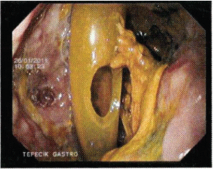
Tube duodenostomy was performed inside the duodenal fistula and duodenostomy was wrapped around with omentum. The operation ended with placing a silicone drainage tube to subhepatic place near the anastomosis. Bilious mai began to drain from both duodenostomy tube and subhepatic drainage tube since the postoperative first day. During postoperative follow-up bilious mai continued draining at the same rate. Gastroduodenoscopy was performed on the postoperative twelfth day. It was found out that the silicone drainage tube was in the duodenum during endoscopic examination (Figure 1). The patient was followed with the diagnosis of iatrogenic duodenal injury. The silicone drainage tube was removed 3 weeks after the operation. Some bilious purulent matter drained from the opening where the silicone tube had been removed for a few days. The duodenostomy tube was removed when the purulent matter stopped draining. The patient had no complication during follow-up and discharged home.

Figure 1: Silicone drainage tube was in the duodenum during endoscopic examination.
Discussion
Gall bladder diseases are mainly consist of clinical findings based on gall bladder stones and their complications and their current treatment is surgery. Gall bladder stones become symptomatic with colicky abdominal pains. Acute cholecystitis occurs by the inflammation of gall bladder and is treated either conservatively or surgically. In acute cholecystitis serous material in the gallbladder becomes infected and bacterias reproduce leading to empyema. Feeding of the gall bladder wall is disrupted leading to necrosis and perforation. Leakage of bile into the abdomen form the perforated bladder makes treatment harder [4,5].
The surgical treatment of gall bladder diseases is either open or laparoscopic cholecystectomy. Partial cholecystectomy, subtotal cholecystectomy, near total cholecystectomy or cholecystostomy are alternative surgical treatment strategies in case of impossibility to perform cholecystectomy. In case of impossibility to perform total cholecystectomy as in our case, gall bladder and infection can re-occur and acute cholecystitis happens again. In this case, the morbidity and mortality rates may become higher [6,7].
We believe that performing elective complementary cholecystectomy before a complication exists in cases whom total cholecystectomy wasn’t performed is more advantageous and causes less complications. We prefer 3 months interval for our cases for elective complementary cholecystectomy. Surgical treatment of complications in patients whom complementary cholecystectomy wasn’t performed tend to cause many problems. What should be done in these situations? Should complementary cholecystectomy be performed or should cholecystostomy or near total cholecystectomy be the choice of treatment? Our answer is in acute cholecystitis cases such as ours’, an experienced surgeon should perform the operation and the least harmful and curative prosedure must be chosen. We recommend that in the absence of adhesions complementary cholecystectomy must be tried but if there are dense adhesions and if separation of these adhesions may cause complications,near total cholecystectomy can be performed. The small remnant gallbladder neck is oversewn in near total cholecystectomy. But if there’s the thought of causing complications with near total cholecystectomy, cholecystostomy can be a temporary solution [8-10].
In our case, complication occured after partial cholecystectomy without performing elective complementary cholecystectomy and cholecystitis with empyema developped. The opration was performed as we explained above but the patient removed the drainage tube accidentally. If we criticize ourselves retrospectively; fixing the drainage tubes to the skin must be done more carefully and stronger since they are vital for patient follow-ups. Additional fixing sutures must be put to drainage tubes that fistula material drains in order to to strenghten the holding of drainage tube to the skin especially in cases that fistulas develop. And patients must be informed about the importance of the drainage tubes and they must be told to take more care.
Our case whom was reoperated with the diagnosis of duodenal fistula, was placed duodenostomy tube and before closing the abdomen subhepatic drainage tube was placed close to the suture line. But the drainage tube involuntarily went into the duodenum by disrupting the suture line. Draining of the same amount and same characteristics of fluid from both tubes on the first postoperative day confused us a lot and made us think that duodenal fistula re-occured again from the first day. It didn’t come to mind that both of the tubes are in the duodenum since it’s a very rare complication. It’ normal not to think of this situation since this hasn’t happened before. If we criticize this situation retrospectively; draining of the same amount and same characteristics of fluid from both tubes means both of the tubes are draining the same place and there’s no barrier between the tubes. In such situations, either the fistula should re-occur or two tubes should come together. Imaging studies can be useful to prove this [11,12].
Our patient was diagnosed with upper gastrointestinal system endoscopy by seeing the silicone drainage tube in the duodenum. When should the drainage tube be removed? Duodenum is an organ far away from the abdominal wall. To remove the drainage tube, it’s waited for the organisation of the surroundings of the drainage tube and epitelization of the drain tract. The recommended time for duodenum is about 3 weeks. When the drainage tube is removed after 3 weeks, the effluence flows out the abdomen from the drain tract without accumulating ( fluid collection) since the tract doesn’t close immediately. The drainage tube is removed in two ways.?n the first way, the drainage tube is totally removed. In the second way, drainage tube is removed 1 or 2 cm every 1 or 2 days until it’s totally removed. The aim of the second way is to aviod the immediate collapse of the drain tract in order to prevent fluid collection. We waited for 3 weeks in our case and we removed the drainage tube totally since we believed that the tract was epithelialized so that it wouldn’t collapse. If we had believed that there wasn’t enough epithelialization and the tract would collapse after removing the drainage tube, would have remove the tube 1-2 cm every 2-3 day intervals.
There were both duodenostomy tube and silicone drainage tube inside the duodenum in our patient. Which one should be removed first? Could all the tubes be removed at the same time? We didn’ ever think of removing two of them at the same time. We believe that removing them at different times will be more effective. Duodenostomy tube was our treatment strategy and we were sure about its structure. The iatrogenic one was a traumatic event that was out of our control and we couln’t be sure of its structure. Since we trust the structure of duodenostomy tube more, we left it last one to remove and we removed the iatrogenic silicone tube first. Some mai draned from skin after removal of silicone tube for 5-6 days and then it stopped draining. After that, duodenostomy tube was removed and full recovery was obtained 5-6 days later. Our patient was discharged in a healthy way. We also want to emphasize that drainage tubes should be placed at a sufficient distance from anastomosis in order to avoid iatrogenic anastomosis perforations and organ injuries.
7097
References
- Salman B, Akin M, Tezcaner T, Az?l?, Utku, et al. (2008)Laparoskopikkolesistektomidenaçikkolesistektomiyedönülenhastalardapreoperatif risk faktörleriveintraoperatifnedenler: 536 hastaninretrospektifanalizi. Gazi Tip Dergisi 19: 60-65.
- Lindenauer SM (1973) Surgical treatment of bile duct strictures.Surgery 73: 875-880.
- Hillis TM, Westbrook KC, Caldwell FT, Read RC (1977) Surgical injury of the common bile duct.Am J Surg 134: 712-716.
- Ibrahim S, Hean TK, Ho LS, Ravintharan T, Chye TN, et al. (2006) Risk factors for conversion to open surgery in patients undergoing laparoscopic cholecystectomy.World J Surg 30: 1698-1704.
- Rifki Jai S, Lakhloufi A, Hidraoui K, Khaiz D, Chehab F, et al. (2004) [Situations of conversion during laparoscopic cholecystectomy: series of 300 cholecystectomies].Tunis Med 82: 344-349.
- Pavlidis TE, Marakis GN, Ballas K, Symeonidis N, Psarras K, et al. (2007) Risk factors influencing conversion of laparoscopic to open cholecystectomy.J LaparoendoscAdvSurg Tech A 17: 414-418.
- Sanabria JR, Gallinger S, Croxford R, Strasberg SM (1994) Risk factors in elective laparoscopic cholecystectomy for conversion to open cholecystectomy.J Am CollSurg 179: 696-704.
- [No authors listed] (1993) Guidelines for the treatment of gallstones. American College of Physicians.Ann Intern Med 119: 620-622.
- Ransohoff DF, Gracie WA (1993) Treatment of gallstones.Ann Intern Med 119: 606-619.
- Kolla SB, Aggarwal S, Kumar A, Kumar R, Chumber S, et al. (2004) Early versus delayed laparoscopic cholecystectomy for acute cholecystitis: a prospective randomized trial.SurgEndosc 18: 1323-1327.
- Sharma AK (2001) External biliary fistula.Trop Gastroenterol 22: 163-168.
- Ramirez MA, Santillana M, Florian M (1988) Use of an elemental diet for nutritional management of severely undernourished and immunocompromised patients with gastrointestinal fistulas: experience in Peru. Nutrition 4: 367m


