Case Report - (2022) Volume 10, Issue 2
IgA-IgA Biclonal Gammopathy of Undetermined Significance: A Case Report and Review of the Literature
Ibtissam MHIRIG*,
Sara Harrar,
Ayoub Bouchehboun,
Saliha Chellak and
Abderrahman Boukhira
Biochemistry Laboratory, Faculty of Medicine and Pharmacy, Cadi Ayyad University, Avicenna Military Hospital, Marrakech, Morocco
*Correspondence:
Ibtissam MHIRIG, Biochemistry Laboratory, Faculty of Medicine and Pharmacy, Cadi Ayyad University,
Morocco,
Email:
Received: 15-Dec-2021, Manuscript No. IPACLR-21-12013;
Editor assigned: 17-Dec-2021, Pre QC No. IPACLR-21-12013(PQ);
Reviewed: 03-Jan-2022, QC No. IPACLR-21-12013;
Revised: 07-Feb-2022, Manuscript No. IPACLR-21-12013(R);
Published:
14-Feb-2022, DOI: 10.36648/2386-5180.22.10.395
Abstract
Biclonal gammopathy is a rare disease entity characterized by the simultaneous appearance of two bands, at different positions, on the Serum Protein Electrophoresis (SPEP) and serum immunofixation electrophoresis (SIFE), resulting from the clonal expansion of two dissimilar neoplastic cell lines. We report a case of a biclonal gammopathy of undetermined significance. SPEP showed a biclonal appearance in the gamma region. SIFE confirmed the presence of an IgA-IgA double band. This combination of heavy chains is rarely described in the literature.
Keywords
IgA-IgA Biclonal gammopathy, Undeteremined significance, Serum
protein electrophoresis, Serum immunofixation electrophoresis.
Introduction
Bi-clonal gammopathy is a rare disease entity characterized by
the simultaneous occurrence of two bands, at different positions,
on the SPEP and/Or SIFE, resulting from clonal expansion of
two dissimilar neoplastic cell lines. It accounts for 3-4% of
all gammopathies. It is associated with lymphoproliferative
syndromes, multiple myeloma or monoclonal gammopathies
of undetermined significance (MGUS), also called a bi-clonal
gammopathy of undetermined significance (BGUS) [1-3].
Several heavy chain isotype combinations have been reported
in the literature [3]. The most commonly observed are: IGGIGA,
IGG-IGG, IGG-IGM, and unusually, a double band IGAIGA.
Thereupon, we report the case of a BGUS with IGA- IGA/
Kappa.
Case Report
75-year-old man, with medical history of hypertension.
He presented to the clinic with asthenia, progressive
lethargy, and chronic gastric pain. The clinical examination
revealed a mucocutaneous pallor, with no other associated
clinical abnormalities, in particular no adenopathies or
hepatosplenomegaly. The blood count showed an iron deficiency
anemia of 9.3 g/dL and a ferritin level of 9.95 ng/ml. The other
leukocyte and platelet cell lines were normal. The blood smear
showed anisopoikilocytosis, hypochromia and target red blood cells. Furthermore, no malignant cells were detected.
The C-reactive protein was 0.48 mg/l. The sedimentation
rate was 11 mm/hour. Liver, renal, albumin and protid levels
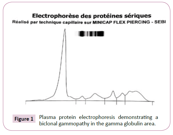
were normal. Plasma protein electrophoresis performed by
capillary electrophoresis technique (MINICAP FLEX PIERCING;
SEBIA®), revealed the presence of a biclonal gammopathy in the
gammaglobulin area without any related hyperglobulinemia.
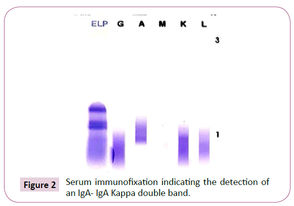
Serum immunofixation was realised on a semi-automatic device
(HYDRASYS; SEBIA®) using an agarose gel as migration support.
It showed the presence of two bands in the IGA migration zone
and in the Kappa zone. Bence Jones proteinuria was normal.
The serum free light chain ratio was 2.16. The myelogram was
normal as well as the standard radiography. Colofibroscopy
performed for the digestive symptomatology did not show any
sign of progressive neoplasia but found a chronic gastritis with
Helicobacter Pylori. The patient received a Helicobacter pilory
eradication therapy, and martial therapy for his iron deficiency
anaemia (Figure 1 and Figure 2).

Figure 1. Plasma protein electrophoresis demonstrating a biclonal gammopathy in the gamma globulin area.

Figure 2. Serum immunofixation indicating the detection of an IgA- IgA Kappa double band.
The diagnosis of biclonal MGUS was retained with strict shortterm
monitoring
Discussion
Biclonal gammopathies involves the occurrence of 2 or more
M proteins in serum and/or urine, which may be composed of
equal or differing combinations of heavy and light chains [4].
The production of these paraproteins can be attributed to the
existence of a single clone of B lymphoid cells, which then diversifies
into 2 independents clones after a process of antigenic selection, or
it is due to the presence of two different clones at the beginning
of tumor transformation [5]. This represents approximately 2.8%
of gammopathies [2]. The most commonly reported cause is
represented by biclonal gammopathies of undetermined significance,
followed by multiple myeloma, Waldenström macroglobulinemia
and other malignant lymphoproliferative disorders [6].
The average age of discovery varies widely from 63 to 72 years
according to the studies [2,7-9]. Male predominance is broadly
described [8-10].
A retrospective study by Ghammad et al. [7] reported that 100%
of patients diagnosed with BGUS did not present clinical signs or
bone lesions or kidney disease or anemia. Similarly, our patient
was asymptomatic, except for asthenia and mucocutaneous
pallor which were explained by his iron deficiency anemia, and
which resolved after introduction of martial therapy.
All associations of immunoglobulin heavy chains and/or light
chains are well described in the literature. According to the study
by Sharma et al, the most common combination immunoglobulin
was IgG-IgA (33%) [10]. These same results are consistent with
those of Katzman et al. [11]. Whereas IgG-IgM isotypes were
the most common according to the cohort study [6,7,12,13].
In the study by Garcia et al, the most prevalent combination of
paraproteins was IgG-IgG [2]. BIGUS IGA-IGA was described in
two cases in the study by Guastafierro [14]. The predominance of
Kappa light chains is described in all of the previously mentioned
series of studies, as was the case for our patient. These differences
in composition may be explained by the small number of cases of
BGUS in each series, or the low sensitivity of techniques for the
detection of para proteins.
Recently, the arrival of capillary electrophoresis has provided
an important support in the diagnosis of gammopathies, it is a
very successful technique that allows a very efficient and rapid
separation of serum proteins with an increased sensitivity and a
specificity comparable to agarose gel electrophoresis [14].
In this case, the capillary SPEP technique showed a double peak in
the gamma globulin area which was confirmed by the existence
of a double band on SIFE. In the series by Sharma et al. [10],
SPEP found a double monoclonal band in 80% of BGUS cases,
confirmed by SIFE in 100% of cases. Similarly for Guastafierro
et al. [14], the double component was determined in 23.5%
of the cases via agarose gel electrophoresis and in 100% of
the cases via SIFE. Kyle et al. [1] reported the presence of 2
localized bands on SPEP in 57.9% of cases, and in the other
42.1% of cases, biclonal gammopathies were identified by
SIFE. In cases where SPEP results are negative, the SIFE
technique is widely accepted as a confirmatory test because
of its higher sensitivity, ease of interpretation, and reliability
[10]. Pretreatment with mercaptoethanol is an an expensive
and a simple methods which depolymerizes the multimers and
helps in better distinction of double gammopathies. It can be
used in facilities where IFE is not available. The latter was used
in our case due to its wide availability in our biochemistry
laboratory.
The risk of malignant transformation into multiple myeloma is
similar to that of monoclonal gammopathies of undetermined
significance. It is about 1% [9]. The two clones are probably the
result of random processes. They are both independent and
probably follow their own natural history in parallel.
lastly, the disappearance of the bands obeys regulatory
processes or may be due to additional mutations generated in
the neoplastic cell lines [15].
Conclusion
IGA-IGA biclonal gammopathy is a rare disease, most frequently
combined with GBSI. GBSI has a similar risk of malignant
transformation as monoclonal gammopathy, but further studies
are required to compare the progression of the 2 monoclonal
and biclonal components and to investigate a possible regional
or national population specification.
REFERENCES
- Kyle RA, Robinson RA, Katzmann JA (1981) The clinical aspects of biclonal gammopathies: Review of 57 cases. Am J Med 71: 999‑1008.
Indexed at, Google Scholar, Cross Ref
- García-García P, Enciso-Alvarez K, Diaz-Espada F, Vargas-Nuñez JA, Moraru M, et al. (2015) Biclonal gammopathies: Retrospective study of 47 patients. Rev Clin Esp 215: 18-24.
Indexed at, Google Scholar, Cross Ref
- Khalid A, Shahbaz I (2018) Double Gammopathy of Undetermined Significance-A First Case Report from Pakistan. J Hematol Thrombo Dis 6: 297.
Google Scholar
- Tschumper RC, Dispenzieri A, Abraham RS, Henderson KJ, Jelinek DF (2013) Molecular analysis of immunoglobulin genes reveals frequent clonal relatedness in double monoclonal gammopathies. Blood Cancer J 3: e112.
Indexed at, Google Scholar, Cross Ref
- Brito-Zerón P, Retamozo S, Gandía M, Akasbi M, Pérez-De-Lis M, et al. (2012) Monoclonal gammopathy related to Sjögren syndrome: a key marker of disease prognosis and outcomes. J Autoimmun 39: 43-48.
Indexed at, Google Scholar, Cross Ref
- Bora K, Das U, Barman B, Ruram AA (2017) Monoclonal gammopathy with double M-bands: An atypical presentation on serum protein electrophoresis simulating biclonal gammopathy. Indian J Pathol Microbiol 60: 590-592.
Indexed at, Google Scholar, Cross Ref
- Ghammad W, Berrada S, Aissaoui M, Slaoui A, Iraqui FZ, et al. (2020) Biclonal Gammopathies: A Retrospective Study in Hassan II University Hospital Center, Fez, Morocco. Sch Int J Biochem 3: 226-231.
- Leroy H, Decaux O, Ianotto JC, Guenet L, Ruelland A, et al. Caractéristiques cliniques et biologiques des gammapathies biclonales. Description d’une cohorte de 203 patients. La Revue De Médecine Interne 29.
Google Scholar
- Mullikin TC, Rajkumar SV, Dispenzieri A, Buadi FK, Lacy MQ, et al. (2016) Clinical characteristics and outcomes in biclonal gammopathies. Am J Hematol 91: 473‑475.
Indexed at, Google Scholar, Cross Ref
- Sharma S, Gupta P, Aggarwal R, Malhotra P, Minz RW, et al. (2019) Demystifying Biclonal Gammopathy: A Pathologist’s Perspective. Lab Med 50: 357-363.
Indexed at, Google Scholar, Cross Ref
- Katzmann J, Kyle RA, Lust J, Snyder M, Dispenzieri A (2013) Immunoglobulins and Laboratory Recognition of Monoclonal Proteins. Neoplastic Diseases of the Blood 565-588.
Google Scholar, Cross Ref
- Riddell S, Traczyk Z, Paraskevas F, Israels LG (1986) The double gammopathies. Clinical and immunological studies. Medicine(Baltimore) 65: 135-142.
Indexed at, Google Scholar, Cross Ref
- Nilsson T, Norberg B, Rudolphi O, Jacobsson L (1986) Double gammopathies: incidence and clinical course of 20 patients. Scand J Haematol 36: 103‑106.
Indexed at, Google Scholar, Cross Ref
- Guastafierro S, Ferrara MG, Sica A, Parascandola RR, Santangelo S, et al. (2012) Serum double monoclonal components and hematological malignancies: Only a casual association? Review of 34 cases. Leuk Res 36: 1274-1277.
Indexed at, Google Scholar, Cross Ref
- Bast EJEG, Slaper-Cortenbach CM, Verdonck LF, Samson W, Ballieux RE, et al. (1985) Transient expression of a second monoclonal component in two forms of biclonal gammopathy. Br J Haematol 60: 91-97.
Indexed at, Google Scholar, Cross Ref
Citation: Ibtissam M, Harrar S, Bouchehboun S, Chellak S, Boukhira A (2022) IgA-IgA Biclonal Gammopathy of Undetermined Significance: A Case Report
and Review of the Literature. Ann Clin Lab Res. Vol.10 No.2:395.


