Allam Fayez Abuhamda1*, Shady Abu El Ajeen2, Wesam Shaltout2 and Salama Abu Nada3
1Gaza Strip’s Neonatal Intensive Care Units, Gaza, Palestine
2Al-Aqsa Neonatal Intensive Care Unit, Al-Aqsa martyrs Hospital, Gaza, Palestine
3Al-Aqsa Martyrs Hospital, Gaza, Palestine
*Corresponding Author:
Allam Fayez Abuhamda
MOH Senior Consultant Neonatologist
Gaza Strip’s Neonatal Intensive Care Units, Gaza, Palestine
Tel: +00972597502720
E-mail: allam570@yahoo.com
Received Date: September 1, 2018; Accepted Date: September 15, 2018; Published Date: September 17, 2018
Citation: Abuhamda AF, Ajeen SAE, Shaltout W, Nada SA (2018) Imperforate Hymen with Hydrocolpos in Palestinian Neonate: A Case Report. Ann Clin Lab Res Vol.6 No.3:255. DOI: 10.21767/2386-5180.100255
Keywords
Neonatal hydrocolpos; Lower urinary tract; Renal failure
Introduction
Imperforated hymen incidence is 0.014-0.01% [1]. The presentation mostly late with amenorrhea at the adolescence age. Specific causes for the failure to set up patency are not clear. The cause may be related to failure of apoptosis due to a genetically transmitted signal, or it may be related to an inappropriate hormonal milieu [2]. Familial inheritance in successive generations has been described [3]. Hydrocolpos is an accumulation of reproduction gland secretion and cystic dilation of the vagina due to imperforate hymen or congenital anomalies of the vagina like; vaginal atresia or membranous obstruction in the lower part of the vagina [4-7]. If the condition not diagnosed early, could be renal failure develops due to lower renal obstruction. Our case is female, she was actually diagnosed to have pelvic-abdominal mass, at birth by examination; she had abdominal distention and imperforate hymen.
Case Report
Second-degree consanguineous parents, they had no family history of congenital anomalies. Mother, she had good antennal care, at 39 weeks gestation age, antenatal ultrasound showed the fetus had pelvic-abdominal mass 4 × 6 cm (Figure 1).
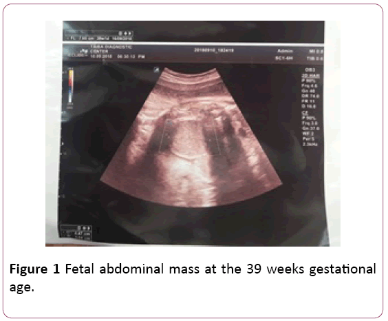
Figure 1: Fetal abdominal mass at the 39 weeks gestational age.
She was born at 40 weeks gestational age, by normal vaginal delivery, Apgar score were 7, 9 at 1, 5 minute respectively. Birth weight was 3300 grams and head circumference was 36 cm. She had abdominal distension in the lower part, by palpation, there is a tender mass with a diameter of about 6 cm (Figure 2). The other parts of the physical examination is normal. She has a normal patent anus and she passed stool. she has a patent normal urethral opening and passed urine 3 ml/kg/day. She had imperforated bulging hymen (Figures 3-5). Abdominal ultrasound showed cystic abdominal mass measure about 5 × 8 cm, there were left-sided hydronephrosis and hydro ureter.
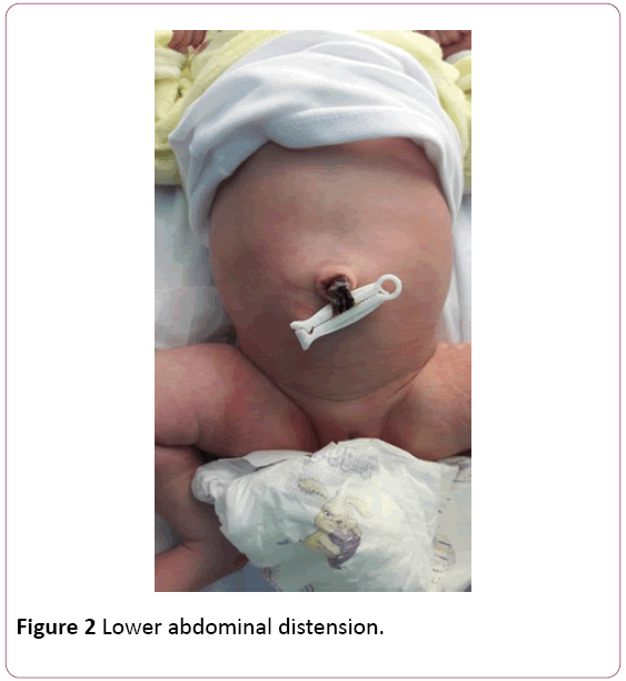
Figure 2: Lower abdominal distension.
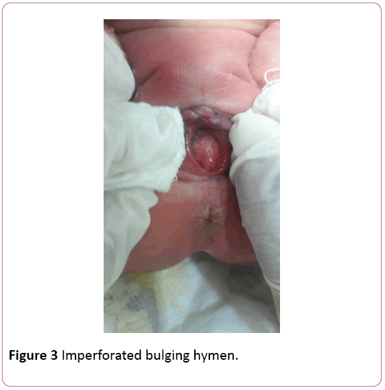
Figure 3: Imperforated bulging hymen.
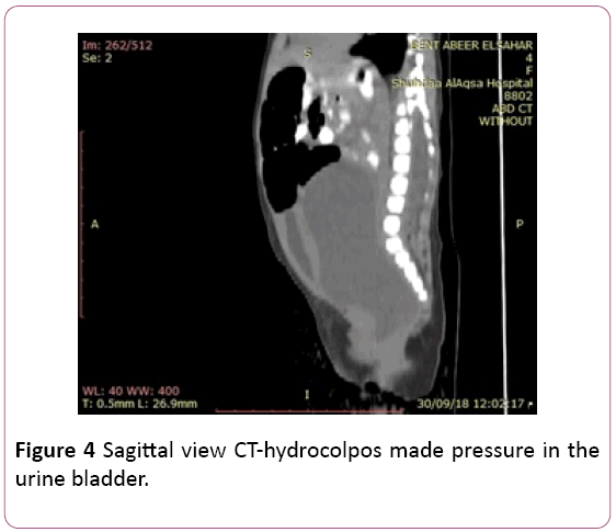
Figure 4: Sagittal view CT-hydrocolpos made pressure in the urine bladder.
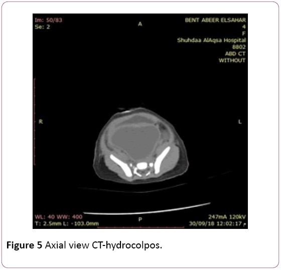
Figure 5: Axial view CT-hydrocolpos.
Abdominal CT showed cystic mass extended from the pelvis the level of the hymen, the mass was not connected to the internal organ or spine. The mass made pressure on urine bladder which was pushed anteriorly. Clinical presentation and abdominal CT was diagnostic of imperforate hymen with hydrocolpos. The pediatric surgeon did a hymeneal incision and a large amount of milky secretion was drained. The abdomen distension significantly subsided. The baby was discharged home to be operated next week under general anesthesia for appropriate vaginoplasty.
Investigation
Serum urea-20 mg/dl; Creatinine-0.3 mmol/l; Sodium-135 mmol/l; Potassium-4 mmol/l; Calcium-1.14 mmol/l.
Discussion
By antenatal ultrasound, the fetus had lower abdominal mass at the age of 39 weeks gestation. By physical examination after birth, she had visible abdominal distension, abdominal ultrasound showed pelvic cystic mass not connected to the internal organ but had pressure on the left side causing hydro-ureter and hydronephrosis, abdominal CT showed cystic mass extended from pelvis to lower part of vagina till hymen. The CT showed also cystic mass made pressure on the urine bladder which was pushed anteriorly. Physical examination also showed bulging imperforated hymen. The clinical examination and the radiologic findings were diagnostic of neonatal imperforated hymen and hydrocolpos. The diagnosis at the time and the appropriate management of pediatric surgeon by hymeneal incision saved the baby from a fatal complication [8,9].
Conclusions
• The genital area of the female new-born should be examined well after birth.
• Early diagnosis and at appropriate management will protect the patient from a fatal complication.
Acknowledgment
To the Dr. Ahmed Shaltout, consultant radiologist, CT department, Al-Aqsa Martyrs Hospital, MOH, Gaza, Palestine.
Conflict of Interest
The authors declare no conflict of interests.
23441
References
- Taneh H, Shahmiri M, Kouchaki GM, Royani R, Chehregosha M, et al. (2017) Imperforate hymen with hydrocolpos: A case report. JCBR 1(3): 20-24.
- Ercan CM, Karasahin KE, Alanbay I, Ulubay M, Baser I (2011) Imperforate hymen causing hematocolpos and acute urinary retention in an adolescent girl. Taiwan J Obstet Gynecol 50(1): 118-120.
- Stelling JR, Gray MR, Davis AJ, Cowan JM, Reindollar RH (2000) Dominant transmission of imperforate hymen. Fertil Steril 74(6): 1241-1244.
- Ramphul M, Perry L, Bhatia C (2016) Neonatal imperforate hymen with hydrocolpos. BMJ case reports.
- Al-Salem, Ahmed H (2017) Hydrocolpos, vaginal agenesis and atresia: An illustrated guide to pediatric urology. Springer Cham pp: 619-633.
- Mistry A, Bandhu S, Davies E, Medd N (2017) What’s that abdominal mass?. Archives of Disease in Childhood-Education and Practice 2: 1.
- Pierluigi Marzuillo, Stefano Guarino, Andrea Apicella, Angela La Manna (2017) Imperforate hymen. Turk J Urol 43(1): 102.
- Vitale V, Cigliano B, Vallone G (2013) Imperforate hymen causing congenital hydrometrocolpos. J Ultrasound 16(1): 37-39.
- Eksioglu AS, Maden HA, Cinar G, Tasci Yildiz Y (2012) Imperforate hymen causing bilateral hydroureteronephrosis in an infant with bicornuate uterus. Case Rep Urol p: 102683.


