Omer Engin1*, Mebrure Evnur Uyar2, Chaitali Sen1, Tore Oguzhan Sunamak3
1Surgery Department, Buca Seyfi Demirsoy State Hospital, Izmir, Turkey
2Emergency Department, Buca Seyfi Demirsoy State Hospital, Izmir, Turkey
3Surgery Department, Haydarpasa Numune Training and Research Hospital, Istanbul, Turkey
- *Corresponding Author:
- Omer Engin, MD
Buca Seyfi Demirsoy State Hospital
Surgery Department <
Izmir, Turkey
E-mail: omerengin@hotmail.com
Keywords
Short bowel syndrome, Short bowel syndrome nutrition, Feeding jejunostomy, Hernia, Bowel resection.
Introduction
In many patients whom laparotomy was performed intra abdominal adhesions can occur in the postoperative period. These adhesions are usually asymptomatic but they can also cause various clinical symptoms. The clinical findings vary from insignificant clinical symptoms to serious and mortal complications. Adhesions can cause mechanical intestinal obstructions. These obstructions can occur at any level of small and large bowel. In our case, internal herniation has occurred as a result of adhesion after appendectomy. We present our case whom chain of events has happened resulting in small bowel necrosis
Case Report
A 42 years old male patient presented to the emergency department with the complaints of abdominal pain, nausea and vomiting. In his past medical history, appendectomy was performed 5 years ago. He had severe abdominal pains time to time for the last two months and this time his pain began two days ago. Before this time he did not apply any hospital. He gave the history of having eaten fish two days ago and he was believing that his pain was related to food intoxication. He had a colicky abdominal pain and he had no discharge of gas or stool. His vital signs were within normal limits. He had rebound tenderness on abdominal examination and the physical examination of other systems were within normal limits. On plain abdominal radiograph there were air-fluid levels. Computed tomography scans showed distension of small intestines and free fluid in the abdomen. Leukocytosis was detected on complete blood count. The patient went under surgery with the diagnosis of mechanical intestinal obstruction and acute abdomen. When the abdomen was opened, serohemorrhagic free fluid was seen. Some parts of small intestines were dilated and necrotic. The adhesion (Figure 1) between intestinal loop and cacum was detected on exploration. A tunnel (Figure 2) was formed between the intestinal loop and bowel mesentery, small intestinal loops were passing through the tunnel forming internal herniation. Mechanical intestinal obstruction was because of internal herniation so that venous blood return was disrupted leading to necrosis and serohemorrhagic free fluid in abdomen. Intestinal loops turned back to their original position after adhesiolysis. There was necrosis in the 180 cm part of small intestine 150 cm away from the Treitz ligament. This part of small intestines were resected and jejunostomy was performed to the proximal part. There was 50 cm part left on the distal part to the cecum and feeding ileostomy was performed to this distal part (Figure 3). In the postoperative period we started orally feeding with enteral nutrition solutions. In addition enteral nutrition solutions were given through feeding ileostomy. The patient was then discharged with the appointment 3 months later for the closure of jejunostomy and feeding ileostomy.
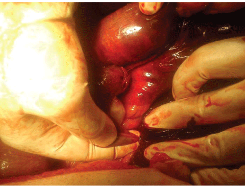
Figure 1: Adhesion between intestinal loop and cacum.
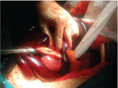
Figure 2: Tunnel was formed between the intestinal loop and bowel mesentery
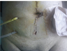
Figure 3: ejunostomy and feeding ileostomy
Discussion
Adhesions are one of the most frequent complications after abdominal surgery.They can be asymptomatic or can cause various symptoms in the early or late postoperative period. Mechanical intestinal obstruction can occur due to these adhesions. Obstructions may regress with medical treatment or operative procedure may be needed. Adhesiolysis is performed for adhesions. Adhesiolysis is needed surgical skills. Adhesiolysis must be made with blunt and sharp dissection. If any serosal injury is occurred, this site must be sutured for prevention of perforation and fistula.
Resection is performed if there is intestinal necrosis. Anastomosis can be performed after resection but if the abdomen is infected, intestinal walls are edematous then, with the thought of postoperative anastomosis leakage, ileostomy or colostomy can be chosen [1-3].
Nausea and vomiting are the most common symptoms in patients with intestinal obstruction. The contents of vomit vary according to the level of the obstruction. The vomit is bilious if the obstruction is in the proximal part of small intestines but contains ileal contents if the obstruction is in t`he distal part of small intestines. If there’s colonic obstruction and ileocaecal valve insufficiency fecaloidal vomiting occurs. If ileocaecal valve is intact and there’s no regurgitation of colonic contents to ileum, then closed loop occurs and pressure in the colon increases leading to colon perforation [4-6].
On plain abdominal radiograph, air-fluid levels are detected and obstruction level can be estimated due to the localization of intestinal gases. In the absence of colonic gas shadow on the plain radiography we may say that the obstruction level is in the small intestines. Also the computed tomography helps us in the diagnosis of obstruction, detection of free fluid and air. It also helps the clinician to determine the localization of tumors if the obstruction is due to malignancy [7,8].
On physical examination there can be tenderness, guarding and rebound parallel to the severity of obstruction. Metallic sound can be heard on auscultation. Necrosis of intestine, perforation, sepsis, free fluid and air are among findings related to the severity of intestinal obstruction [9].
There was an adhesion between the intestinal loop and cecum and a tunnel occurred between intestinal loop and bowel mesentery in our patient. Strangulation has occurred with the intestines passing through the tunnel. We believe the origin of unexplained abdominal pain over two months period was internal herniation. When we think retrospectively, MR enteroclysis or small intestine barium study would help to diagnose. These imaging tests could be prevented from short bowel syndrome [10,11].
Since the abdomen was infected and intestinal edema and inflammation occurred, anastomosis after resection wasn’t performed and enterostomy and feeding ileostomy were preferred. Since the colon and distal 50 cm part of ileum were intact, feeding ileostomy was performed in order to continue the function of absorption. We believe that feeding ileostomy is important for our patient since proximal intestine is short and absorption will decrease.
6019
References
- Ellis H (1997) The clinical significance of adhesions: focus on intestinal obstruction.Eur J SurgSuppl : 5-9.
- Fevang BT, Fevang J, Lie SA, Søreide O, Svanes K, et al. (2004) Long-term prognosis after operation for adhesive small bowel obstruction.Ann Surg 240: 193-201.
- Kuijpers HC, Klok S (2014) Prevention and Treatment of Postoperative Complications after Stoma Surgery. In: Treatment of Postoperative Complications after Digestive Surgery. Cuesta MA, Bonjer HJ (eds), Springer London.
- Jackson PG, Raiji MT (2011) Evaluation and management of intestinal obstruction.AmFam Physician 83: 159-165.
- Chalya PL, Mabula JB, Chandika AB (2014) Dynamic bowel obstruction: aetiology, clinical presentation, management and outcome at Bugando Medical Centre, Mwanza, Tanzania. Tanzania Journal of Health Research16: 1.
- Sahoo MR, Kumar A, Jaiswal S, C B (2013) Transverse colon perforation due to carcinoma rectum: an unusual presentation against Laplace's law.BMJ Case Rep 2013.
- Minordi LM, Vecchioli A, Larosa L, (2012) Radiological Diagnosis of Small-Bowel Diseases. In: Ileoscopy.Trecca A (eds), Springer Milan.
- Hayakawa K, Tanikake M, Yoshida S, Yamamoto A, Yamamoto E, et al. (2013) CT findings of small bowel strangulation: the importance of contrast enhancement.EmergRadiol 20: 3-9.
- Hucl T (2013) Acute GI obstruction.Best Pract Res ClinGastroenterol 27: 691-707.
- Frager D, Baer JW, Medwid SW, Rothpearl A, Bossart P (1996) Detection of intestinal ischemia in patients with acute small-bowel obstruction due to adhesions or hernia: efficacy of CT.AJR Am J Roentgenol 166: 67-71.
- Fletcher JG, Fidler JL,HuprichJE (2012) Small-bowel imaging with CT and MR: Overview of techniques and indications. Applied Radiology41: 18.

