Jonette M Bartlett, Thanh D Hoang* and Alfred F Shwayhat
Department of Internal Medicine, Division of Endocrinology, Naval Medical Center, San Diego, USA
- *Corresponding Author:
- Thanh D Hoang
Department of Internal Medicine
Division of Endocrinology
Naval Medical Center
San Diego, USA
E-mail: Thanh.Hoang@med.navy.mil
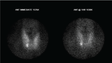
A 44 year-old female presented with one-year-history of fatigue, irritability, forgetfulness, and esophageal reflux. She was taking lisinopril for hypertension and atorvastatin for hyperlipidemia. She had no pertinent family medical history. Physical examination was normal. Laboratory results: serum calcium 11.0 (8.9-10.3 mg/dL), albumin 3.8 (3.5-5.0 mg/dL), PTH intact 167 (ref 10-65 pg/mL), phosphate 2.1(2.5-4.6 mg/dL), 25-hydroxy-Vitamin D 11 (30-100 ng/mL). Tc-99m MIBI scan suggested a right inferior parathyroid adenoma (Figure 1A). Bilateral neck exploration failed to identify parathyroid adenoma. Postoperatively PTH 184 pg/ mL, phosphate 2.2 mg/dL, calcium 11.1 mg/dL.
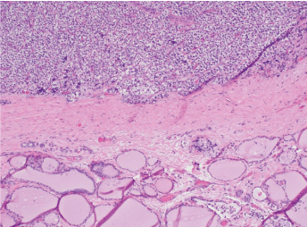
A 44 year-old female presented with one-year-history of fatigue, irritability, forgetfulness, and esophageal reflux. She was taking lisinopril for hypertension and atorvastatin for hyperlipidemia. She had no pertinent family medical history. Physical examination was normal. Laboratory results: serum calcium 11.0 (8.9-10.3 mg/dL), albumin 3.8 (3.5-5.0 mg/dL), PTH intact 167 (ref 10-65 pg/mL), phosphate 2.1(2.5-4.6 mg/dL), 25-hydroxy-Vitamin D 11 (30-100 ng/mL). Tc-99m MIBI scan suggested a right inferior parathyroid adenoma (Figure 1A). Bilateral neck exploration failed to identify parathyroid adenoma. Postoperatively PTH 184 pg/ mL, phosphate 2.2 mg/dL, calcium 11.1 mg/dL. Bedside thyroid ultrasound showed a 9 mm hypoechoic lesion with increased vascularity within the inferior portion of the right thyroid lobe (Figure 1B). Repeat sestamibi scan showed a persistent focus of increased accumulation of radiotracer in the inferior pole of the right thyroid lobe. FNA of this lesion showed lymphocytic thyroiditis with cystic degenerative changes (Figure 2A); and PTH measurement from FNA was 4737 pg/mL. The patient then underwent right thyroid lobectomy with without complications. Postoperative PTH 14.9 pg/mL, calcium 9.4. Pathology showed a cellular proliferation of clear cells encapsulated by a rim of fibrous tissue separated from colloid follicles on the periphery (Figure 2B).

Figure 1A: Right inferior parathyroid adenoma.

Figure 1B: Inferior portion of the right thyroid lobe.

Figure 2A: lymphocytic thyroiditis with cystic degenerative changes.

Figure 2B: Fibrous tissue separated from colloid follicles on the periphery.
What is the Diagnosis?
Answer: Intrathyroidal parathyroid adenoma. FNA of a parathyroid adenoma can mimic cytological features of lymphocytic thyroiditis; however, aspirates of parathyroid adenoma show elevated intact parathyroid hormone measurements [1,2]. It has been reported that cytology and immunocytochemistry may play an important role in the interpretation of US-guided FNA for preoperative localization of parathyroid tissue [3]. If an intrathyroidal parathyroid gland is suspected in a patient with primary hyperparathyroidism an FNA can distinguish adenoma from a thyroid nodule. In this patient, pathology confirmed a 0.6 cm right inferior intrathyroidal adenoma with otherwise benign thyroid disease. This case demonstrated the usefulness of FNA of parathyroid lesion in a patient with primary hyperparathyroidism and an intrathyroidal parathyroid adenoma who had failed prior bilateral neck exploration. It may be important to perform a combined approach of cytology and PTH measurement (immunochemistry) in diagnosing and localizing parathyroid tissue.
Disclaimers
The views expressed in this article are those of the authors and do not necessarily reflect the official policy or position of the Department of the Navy, Department of Defense, or the U.S. Government.
We certify that all individuals who qualify as authors have been listed; that each has participated in the conception and design of this work, the analysis of data, the writing of the document, and the approval of the submission of this version; that the document represents valid work; that if we used information derived from another source, we obtained all necessary approvals to use it and made appropriate acknowledgements in the document; and that each takes public responsibility for it.
7078
References
- Auger M, Charbonneau M, Huttner I (1999) Unsuspected intrathyroidal parathyroid adenoma: mimic of lymphocytic thyroiditis in FNA specimens- A Case Report. Diagnostic Cytopathology 4:276-279.
- Absher KJ, Truong LD, Khurana KK, Ramzy I (2002) Parathyroid cytology: avoiding diagnostic pitfalls. Head Neck 24: 157-164.
- Abati A, Skarulis MC, Shawker T, Solomon D (1995) Ultrasound-guided fine-needle aspiration of parathyroid lesions: a morphological and immunocytochemical approach.Hum Pathol 26: 338-343.


