Bouthouri Abir*, Mansour Malek, Zaouali Jamel and Mrissa Ridha
Department of Neurology, Tunis Military Hospital, Mont Fleury, 1008 Tunis, Tunisia
Corresponding Author:
Bouthouri Abir
Department of Neurology, Tunis Military Hospital
Mont Fleury, 1008 Tunis, Tunisia
Tel: 71562900
E-mail: abirabir1985@gmail.com
Received date: February 04, 2017; Accepted date: February 09, 2017; Published date: February 16, 2017
Citation: Abir B, Malek M, Jamel Z, Ridha M. A Bair’s Crushing Hug: An Unusual Cause of Pseudo pneumothorax. Arch Med. 2017, 9:1. doi: 10.21767/1989-5216.1000195
Copyright: © 2017 Abir B, et al. This is an open-access article distributed under the terms of the Creative Commons Attribution License, which permits unrestricted use, distribution, and reproduction in any medium, provided the original author and source are credited.
Keywords
Ischemic stroke; Granulomatosis
Case Report
Diffusion weighted magnetic resonance imaging (MRI) of the brain showed increased signal in the right lateral region of the medulla oblongata consistent with an infarction (Figure 1). Angio-MRI was normal.
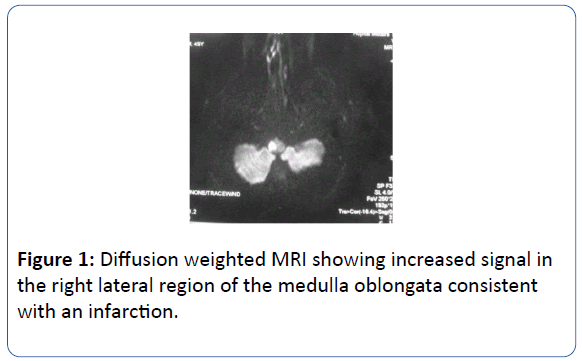
Figure 1: Diffusion weighted MRI showing increased signal in the right lateral region of the medulla oblongata consistent with an infarction.
Laboratory data demonstrated an inflammatory syndrome. Cardiac investigations were normal.
Indirect immunofluorescence tests for cANCA were positive while antinuclear and anti-phospholipid antibodies were negative. Thrombophilia testing was negative.
The patient has been anticoagulted with good clinical outcome.
By one month, the patient presented with sudden bilateral visual loss, fever, respiratory failure associated with necrotic purpura. Glomerular renal impairment appeared.
Chest CT scan disclosed biapical parenchymal densities as well as in the right upper lobe. Pulmonary tuberculosis and lung neoplasm were dismissed.
Ear nose throat examination revealed the presence of polyp on the right maxillary sinus. The retinal angiography showed bilateral thrombosis of the central retinal artery.
The patient was started on intravenous pulse of cyclophosphamide, oral glucocorticoids, courses of immunoglobulins and plasmapheresis for a presumed diagnosis of WG.
There was clinical improvement six months after discharge and cyclophosphamide was switched to azatioprine to maintain remission.
Discussion
This observation illustrates a case of WG whose first manifestation was an ischemic stroke. Our patient satisfies the American College of Rheumatology criteria for WG. He had the classical multisystem picture dominated by lung and kidney involvement, coupled with the positivity of ANCA. The imputability of vasculitis as pathophysiological mechanism of ischemic stroke was established in view of systemic context and an exhaustive etiological investigation involving cardioembolic or atherosclerotic origin, infective endocarditis and essential thrombophilia.WG involves the nervous system in 22% to 54% of cases [1]. Peripheral neuropathy is frequently associated with this disorder.
CNS involvement is rare and occurs in refractory disease. It concerns only 7% to 11% of patients in major series in the literature [2]. It seems to be rarely the mode of entry into the disease, occurring most often late, with an average delay between the onset of symptoms of WG and neurological involvement ranging from five months to more than seven years. It is the initial disease manifestation in 1 to 2.5% of cases [3]. It is usually caused by in situ vasculitis, intracranial granuloma formation or contiguous invasion from extracranial sites.
Intracerebral haemorrhage, seizures, meningeal irritation and mass lesions as a result of granuloma formation are well documented sequelae of WG. Cerebral vessel occlusion by granulomata has also been reported [4]. There have been case reports of ischaemic stroke associated with WG, but generally these are associated with extensive areas of infarction [1]. MRI is a key to diagnosing cerebral vasculitis. Multiple infarcts of varying ages in more than one vascular territory are highly suggestive of an underlying inflammatory process [5]. Cerebral vasculitis may therefore mimic atherosclerotic or hypertensive cerebral events, consequently requiring a high degree of clinical suspicion to make the correct diagnosis. Histological evidence is rarely obtained during brain involvement in WG and the initiation of treatment is then based on a body of clinical and radiological arguments.
There is no specific recommendation for CNS involvement therapy in WG, due to a lack of specific studies [1]. Therapeutic management is still based on the traditional consensual treatment, plasma exchange in addition to the standard induction immunosuppressive regime of cyclophosphamide (or rituximab) plus glucocorticoids [1]. Treatment response is best indicated by an improvement in symptoms rather than resolution of neuroimaging findings, which may remain unchanged. A lack of new lesions developing is highly supportive of treatment response and a follow up MRI should be organized four to six weeks after commencing treatment.
Cerebral vasculitis in WG is a marker of severe disease activity and seems to be associated with a pejorative prognostic impact [3]. Unlike the other cases reported in the literature, our patient, in whom the stroke was made through a cerebral vasculitis, had a favorable outcome with clinical and immunological remission at 30 months of starting treatment.
Conclusion
Stroke is a rare inaugurated manifestation of WG. We suggest that MRI scan should be among standard investigations for a patient presenting with signs of cerebral ischaemia in whom vasculitis is suspected,even in the absence of initial extra neurological manifestations.
18359
References
- Pasqueta F, Karkowskia L, Hajeka V (2012) Une manifestation inaugurale inhabituelle de la granulomatose de Wegener: L'accident vasculaire cerebral ischemique. La Revue de médecine interne 33: 6-9.
- Bares M, Muchova M, Dufek M (2002) Wegener’s granulomatosis: ischemic stroke as the first manifestation (case study). J Neurol. 249: 1593-1594.
- Seror R, Mahr A, Ramanoelina J (2006) Central nervous system involvement in Wegener granulomatosis. Medicine (Baltimore) 85: 54-56.
- Yuan J, Guan HZ, Wu QJ (2004) Neurological manifestations of 33 patients with Wegener’s granulomatosis. Chinese Journal of Neurology 37: 428-430.
- Kuker W, Gaertner S, Nagele T (2008) Vessel wall contrast enhancement-a diagnostic sign of cerebral vasculitis. Cerebrovascular Diseases 26: 23-29.


