Background & aims: This study examines if modelling the cephalocaudal regions into four separate regions reveals different kinematic patterns of spinal motion in relation to hip motion when lifting an object (a 5 kg dumbbell) from the ground to an upright position.
Methods: Thirty-two male participants (mean age = 28.2 ± 4.2 years; weight = 74.4 ± 11 kg; height =1.70 ± 0.04 meters) agreed to participate in this study. The cephalocaudal region of the spine was divided into four distinct regions of the spine (Head-Cervical, Thoracic, Upper-Lumbar and Lower-Lumbar) to obtain their range of motions and velocities against the hip region during a sagittal objectlifting task.
Results: There were significant differences in the range of motions between each of the five regions (p<0.001) during an object-lifting task as well as velocities significant differences between number of spinal regions and hip. The hip region produced the most kinematic motion and velocity followed by the Lower-Lumbar and Upper-Lumbar during the functional lifting task. There were significant correlations found between head-cervical and lower-lumbar, head-cervical and hip, thoracic and upper-lumbar, Thoracic and hip, upper-lumbar and lower-lumbar, and Lower-Lumbar and hip during both range of motion and velocity analysis.
Conclusion: The results of this study demonstrate differences in the contribution of five anatomical regions during a functional lifting task. Hip and Lower-Lumbar and Upper-Lumbar made up the most significant proportion of total kinematic motions and velocity during the functional lifting task.
Keywords
Spine; Range of motion; Velocity; Hip; Cephalocaudal; Sagittal objectlifting
Introduction
Numerous factors cause Lower Back Pain (LBP), one of the most common of which is erroneous object lifting. Indeed, one of the most significant factors that cause LBP among handling workers is poor lifting technique. Therefore, healthcare providers always recommend a proper lifting technique in which the hands are positioned close to the body while lifting objects to avoid spinal injuries. The everyday task that most pertinently influences spine and hip kinematics is lifting objects from the ground, which is a systematic and regular activity especially for persons occupied in physical labour [1]. Commonly, employers provide training sessions for their staff engaged in handling duties to prevent LBP [2]. This training mainly educates them on the proper way to lift objects, as this can place a considerable load on the spine and is regularly mentioned as provoking LBP [3]. Indeed, human spinal Range of Motion (ROM) information is key to developing clinicians’ awareness of spinal disorders [4].
Accurate knowledge of physiological movement of the lumbar spine and hip regions and the behaviour of each regional movement during an object-lifting task is important. Spine and hip motion play an essential role in daily functional activities, such as self-caring or performing occupational duties. The interest of researchers and therapists in movement behaviour has grown, revealing the extent of injuries suffered and also bringing about improvements in movement behaviour. Measuring the regional breakdown of spinal motion in the sagittal plane and describing the relative motion of different regions of the spine can provide useful clinical information, which can be used in clinical procedures for spinal assessment [5]. A considerable amount of literature has been focused on measuring the lumbar spine comparative to hip movement, while performing a number of daily duties [1,6-8]. However, the majority of these studies have treated the lumbar spine as a single region. Over the last ten years, a number of studies have adopted multi-regional lumbar spine models across clinical populations [9,10] and healthy subjects [4,5,11-14], identifying differences in regional contribution. Clinical studies have previously confirmed differences in this ratio between those with and without back pain [1,8], whilst alterations in this ratio affect the bending and compressive stresses on the lumbar spine [15,16]. Subsequently, another study investigated how the upper and lower lumbar regions contributed to spinal movement relative to hip motion, when performing everyday tasks [5]. Unfortunately, study [5] focused on the lumbar spine while the other spinal regions were overlooked in terms of movement over time relative to hip kinematic behaviours. The study of a multi-regional lumbar spine model versus hip motion over time during object lifting has not yet been undertaken. Data arising from such a study would significantly assist in achieving a better understanding of lumbar spine kinematics, especially when supplemented by multiregional velocities [17], as the relative movement behaviour of the hip and its interaction with the lumbar spine is considered important [18,19].
Therefore, this study aimed to examine if modelling the cephalocaudal regions into four separate regions would give a different kinematic pattern of spinal motion with respect to hip motion. This is the first study to adopt this approach to the kinematic analysis of multi-spinal regions against the hip region during an object-lifting task.
Materials and Methods
Participants
Thirty-two male participants (mean age =28.2 ± 4.2 years; weight =74.4 ± 11 kg; height =1.70 ± 0.04 meters) agreed to participate in this study. The Scientific Research Ethical Committee at Najran University approved this study (Ethic no. 442-37-5101 at 22- 01-1442), and its participants were recruited via social media applications which were used to advertise to staff, undergraduate students, and the community surrounding Najran University. In addition, the researcher also issued invitations orally, meaning our cohort was a convenience-based sample. This study faced difficulty in recruiting both genders, particularly females; therefore, all participants in this study were male. Moreover, among those who initially agreed to participate, some of them denied to complete the experimental process when they asked to get their shirts off in order to attach sensors into their bodies. All participants were selected based on specific criteria. Each participant was provided with a sheet containing the study information, indicating their rights, confirming the confidentiality of information, outlining potential risks, and explaining what to do in the event of something going wrong. Each participant had consented to participate. Finally, participants provided written informed consent before participating in this study.
Instrumentation
A six tri-axial accelerometer sensors system was applied on a number of points on each participant’s body. This system was explained in a previously published study [4] and had shown excellent reliability relating to spinal motion analysis with an intraclass correlation coefficient between 0.88 and 0.99 and a standard error of measurement between 0.4° and 5.2°. Moreover, the tri-axial accelerometer sensors system has been used to measure spinal regions and hip kinematics in a number of other studies [4,5,11].
Procedures
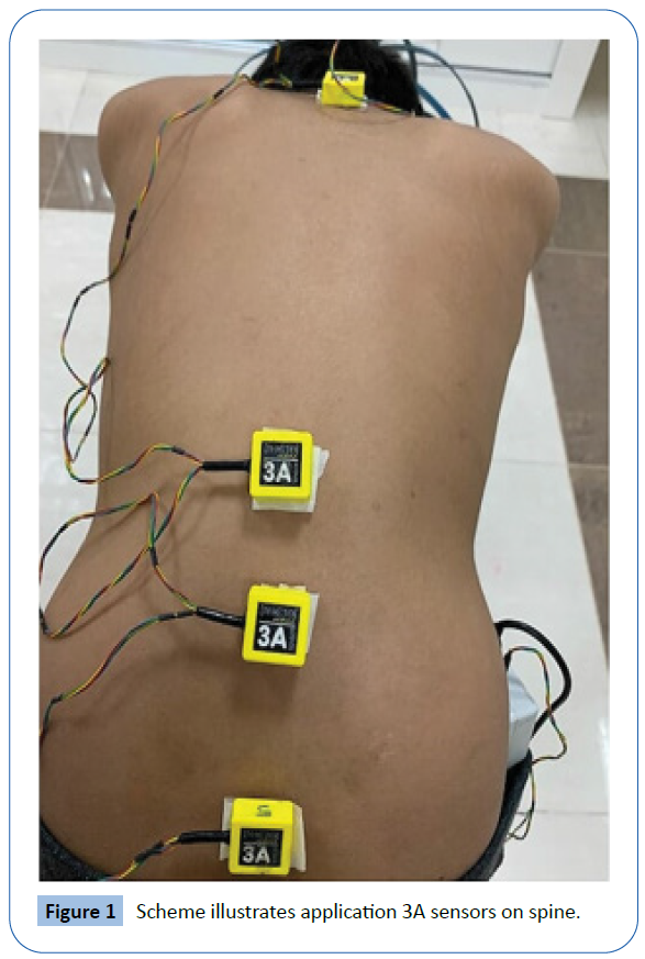
All of the experiments were conducted in the research laboratory of the physiotherapy clinics in the College of Applied Medical Sciences, Najran University. Participants were asked to wear light clothes when it came to measuring their height and weight and to then sit on a chair without a backrest. A medical swab was used to disinfect the places where the sensor needed to be attached. Sensors were affixed using double-sided adhesive tape at six different locations: the forehead, the first and twelfth thoracic vertebra, the third lumbar vertebra, the first sacral vertebra (Figure 1), and the lateral aspect of the mid-thigh. The sensors measured the movement and velocities of the headneck, thoracic, upper and lower lumbar regions associated with hip during the task of lifting an object off the ground (a 5 kg dumbbell (1 pc)) and returning to an upright position. Such an approach was also used by Alqhtani et al. [11] to measure spinal regions over time relatively.
Figure 1: Scheme illustrates application 3A sensors on spine.
Data analysis
Data were collected at 30 Hz and the raw data were relocated to MATLAB (R2011a) and filtered at 6 Hz (low-pass, Butterworth) to delete high-frequency noise [20]. The Head-Cervical (HC) was expressed as the relative angle between the forehead and T1 sensors. The Thoracic region (T) was defined as the relative angle between the T1 and T12 sensors. The Upper Lumbar spine region (UL) was stated as the relative angle between the L3 and T12 sensors, and the Lower Lumbar spine region (LL) as the relative angle between the S1 and L3 sensors. Hip kinematics was determined by the relative angle between the S1 and thigh sensors. Positive and negative velocities of all regions during the performing of an object-lifting task were obtained by distinguishing the ROM data. Each region measurement (either ROM or velocity) was defined as the relative motion between adjacent distal and proximal sensors (i.e., relative angles).
Consequently, regional spinal ROM-time curves were generated for HC, T, UL, LL, and Hip from which peak ROM values [4].
Correlations between regional movements and velocities were explored comparing ROM and velocity profiles using Pearson correlation coefficient calculated in matrix laboratory software (Matlab R2011a, MathWorks). One-way analysis of variance was performed using the Statistical Package for the Social Sciences software (Statistics 21, IBM, Armonk, NY) to determine if significant differences are evident between the region's kinematics. Post hoc analysis was carried out using the Turkey procedure to determine the location of any differences. Statistical significance was accepted at the 5% level for all tests. Spinal regions and hip ROM and velocities ratios were determined for object-lifting task using Excel formulas.
Results
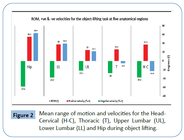
Table 1 details the descriptive statistics for mean (SD) range of motion and velocity for the object-lifting task at five anatomical regions. Where the hip region moved about 63 degrees, the lower and upper lumbar regions moved around 40 and 22 degrees, respectively, during the object-lifting task. However, thoracic (-5 degrees) and head-cervical (-22 degrees) regions moved in an opposite direction. Similarly, the hip region showed the highest positive and negative velocity followed by the LL and H-C regions during the object-lifting task (Figure 2).
| Regions |
ROM (°) |
Positive velocity (°s-1) |
Negative velocity (°s-1) |
| H-C |
-22.8 (19) |
37.3 (17) |
-37.4 (17) |
| T |
-5.4 (14) |
26.4 (14) |
-28.0 (15) |
| UL |
22.1 (9) |
25 (10) |
-22.1 (11) |
| LL |
39.5 (11) |
38.0 (11) |
-37.7 (13) |
| Hip |
63.4 (15) |
55.1 (25) |
-57.9 (26) |
Head-Cervical (H-C); Thoracic (T); Upper Lumbar (UL); Lower Lumbar (LL); Range of Motion (ROM).
Table 1 Mean (SD) range of motion and velocity for the object-lifting task at five anatomical regions.
Figure 2: Mean range of motion and velocities for the Head- Cervical (H-C), Thoracic (T), Upper Lumbar (UL), Lower Lumbar (LL) and Hip during object lifting.
Table 2 presents the differences, correlations, and regional ratios for ROM and velocities of five anatomical regions, revealing significant differences in the ROM during the object-lifting task. The ratio of hip motion was larger than other regional motions during the object-lifting task. Similarly, the ratios of UL and LL motion were greater than the H-C and T regions during the lifting task. Additionally, all of the anatomical regions showed significantly different positive and negative velocities except HC and T, or HC and LL, or T and UL during the object-lifting task.
| Comparative regions |
ROM (p) |
ROM (r) |
Ratio of ROM |
Positive velocity (p) |
Positive Velocity (r) |
Ratio of regional +ve vel |
Negative velocity (p) |
Negative velocity (r) |
Ratio of regional -ve velocity |
| HC T |
0.00 |
0.02 |
0.08 |
0.07 |
0.3 |
1.41 |
0.2 |
0.48 |
1.33 |
| HC UL |
0.00 |
0.28 |
1.29 |
0.03 |
0.02 |
1.49 |
0.004 |
0.16 |
1.69 |
| HC LL |
0.00 |
0.48 |
1.96 |
0.97 |
0.24 |
1.01 |
0.1 |
0.51 |
0.99 |
| HC Hip |
0.00 |
0.17 |
4.18 |
0 |
0.27 |
0.67 |
0.00 |
0.31 |
1.45 |
| T UL |
0.00 |
0.22 |
2.03 |
0.09 |
0.57 |
1.05 |
0.62 |
0.33 |
1.27 |
| T LL |
0.00 |
0.2 |
3.25 |
0.048 |
0.55 |
0.69 |
0.17 |
0.5 |
0.74 |
| T Hip |
0.00 |
0.29 |
3.88 |
0.00 |
0.6 |
0.47 |
0 |
0.51 |
0.48 |
| UL LL |
0.00 |
0.17 |
2.18 |
0.02 |
0.51 |
0.65 |
0.03 |
0.28 |
0.58 |
| UL Hip |
0.00 |
0.04 |
3.57 |
0.00 |
0.37 |
0.45 |
0.00 |
0.39 |
0.38 |
| LL Hip |
0.00 |
0.45 |
1.82 |
0.01 |
0.63 |
0.68 |
0.00 |
0.52 |
0.65 |
Head-Cervical (H-C); Thoracic (T); Upper Lumbar (UL); Lower Lumbar (LL); Range of Motion (ROM)
Table 2 Table demonstrating significant differences (p-value), correlation (r) and regional ratio for ROM and velocity of five anatomical regions.
There were fair correlations between HC and UL or hip, or T and UL or LL or hip, or UL and LL kinematic motion during the object-lifting task. Meanwhile, there was medium correlation between HC and LL, or LL and hip ROM during the object-lifting task. Moreover, there were fair to strong correlations between positive or negative velocities during the object-lifting task over all five anatomical regions.
Discussion
The current study examined the kinematic patterns of spinal and hip motion using four separate regions of the cephalocaudal region. This is the first study to have adopted this approach to examine a kinematics (ROM and velocity) of cephalocaudal and hip regions during an object-lifting task. The results of this study demonstrate significant differences between kinematic motions of separate cephalocaudal regions (HC, T, UL, or LL) and the hip region. The results of this study suggest that separating the cephalocaudal region into four separate regions may help in exploring the contributions of spinal and hip regions during an object-lifting task. A previous study compared two separate lumbar spine regions (UL and LL) with the traditional single unit model. Like the current study, that previous study found that the LL kinematic motion contributed more than other spinal regions during a range of daily activities. Additionally, other studies have also demonstrated different kinematic behaviours of the UL and LL spines, which a traditional single joint lumbar model would fail to recognize [9,13,21].
The current study showed significantly different positive and negative velocities between most of the spinal regions and hip region during an object-lifting task. It indicates the importance of separating the spine into separate anatomical regions when exploring the higher order kinematics (e.g. velocities) of different spinal and hip regions during an object-lifting task. Similarly, a previous study found significant differences between two separate lumbar regions (UL and LL) during the lifting task [1,21]. However, the previous study reported lower velocities of the UL and LL regions during a lifting task than those found in the current study [1,21]. These differences may be due to various methodological factors such as age, sex (both male and female versus male only) and pathological condition (e.g. in pain). Additionally, the current study indicates that the LL region moved at a greater velocity compared to other spinal regions during the object-lifting task. Similarly, the previous study also reported greater velocity of the LL region compared to other regions during a lifting task.
The results of the current study revealed fair to excellent correlations between HC or T or UL or hip and other anatomical regions during the object-lifting task. They also demonstrated fair to moderate correlations between five anatomical regions and positive or negative velocities during the object-lifting task. A previous study reported a strong correlation between UL and LL during a lifting task [14]. The associations between velocity of different spinal and hip regions during an object-lifting task have not been previously investigated. Therefore, the results of this study provide new insights into the relationship between separate regions of the spine and the hip region. While velocity is one of the most important factors with regard to movement quality and may thus give vital kinematic information during an object-lifting task [21], the results of this study have important clinical implications, having offered new information related to velocities of spinal regions and the hip region. A growing body of evidence recommends the assessment of separate spinal regions (e.g. upper and lower lumbar) to identify the role of functionally individual spinal regions in lower back disorders [18,22]. The current study supplements this evidence that the four separate spinal regions are functionally different in terms of ROM and velocity during the object-lifting task and, therefore, assessment of these separate regions could provide a more detailed spinal kinematic model [13]. Greater contributions to motion from the lower lumbar and head-cervical regions, as well as greater movement velocities at these regions, may help to understand the increased prevalence of pathological change in the lower lumbar and cervical regions compared to other spinal regions [23,24]. Furthermore, greater degeneration often takes place in the lower lumbar and cervical regions [25,26] and it is suggested that this is mainly due to greater mechanical stress being placed upon these regions [27].
The current study acknowledges some potential limitations. First, this study included only male participants; therefore, its results cannot be generalized for a whole population. Second, the sample included only asymptomatic young adults. The inclusion of the elderly population or people with pathological disorders (e.g. LBP) might have given different findings. Moreover, the analysis in the current study was limited to the sagittal plane. The use of 3-dimensional analysis would have given detailed kinematics regarding out-of- plane motions.
Conclusion
The novelty of this study is in its examination of the ROM and velocity of four separate spinal regions against the hip region. This is the first study to have adopted this approach to examining the kinematic of multiple spinal regions against the hip region during an object-lifting task. The results of this study demonstrate differences in the contributions of five anatomical regions during a functional lifting task. Hip and lumbar (LL and UL) were responsible for the highest proportion of total kinematic motions and velocity during a functional lifting task. Therefore, physiotherapists should take into account that the lower lumbar spine and hip are more commonly subjected to injuries when they undertake spinal assessment procedures.
As well as it is recommended that future studies in this field be conducted on both males and females of different ages. It is also advised to include LBP sufferers or a mixture of both LBP sufferers and healthy subjects in further research.
References
- Shum GLK, Crosbie J, Lee RYW (2005) Effect of low back pain on the kinematics and joint coordination of the lumbar spine and hip during sit-to-stand and stand-to-sit. Spine 30: 1998-2004.
- Nolan D, O’Sullivan K, Stephenson J, O’Sullivan P, Lucock M (2018) What do physiotherapists and manual handling advisors consider the safest lifting posture, and do back beliefs influence their choice? Musculoskelet Sci Pract 33: 35-40.
- Coenen P, Gouttebarge V, Van Der Burght ASAM, Van Dieën JH, Frings-Dresen MHW, et al. (2014) The effect of lifting during work on low back pain: A health impact assessment based on a meta-analysis. Occup Environ Med 71: 871-877.
- Alqhtani RS, Jones MD, Theobald PS, Williams JM (2015) Reliability of an accelerometer-based system for quantifying multiregional spinal range of motion. J Manipulative Physiol Ther 38: 275-281.
- Alqhtani RS, Jones MD, Theobald PS, Williams JM (2016) Investigating the contribution of the upper and lower lumbar spine, relative to hip motion, in everyday tasks. Man Ther 21: 268-273.
- Lee RYW, Wong TKT (2002) Relationship between the movements of the lumbar spine and hip. Hum Mov Sci 21: 481-494.
- Wong TKT, Lee RYW (2004) Effects of low back pain on the relationship between the movements of the lumbar spine and hip. Hum Mov Sci 23: 21-34.
- Shum GLK, Crosbie J, Lee RYW (2007) Movement coordination of the lumbar spine and hip during a picking up activity in low back pain subjects. Eur Spine J 16: 749-758.
- Williams JM, Haq I, Lee RY (2012) Dynamic lumbar curvature measurement in acute and chronic low back pain sufferers. Arch Phys Med Rehabil 93: 2094-2099.
- Williams JM, Haq I, Lee RY (2013) The effect of pain relief on dynamic changes in lumbar curvature. Man Ther 18: 149-154.
- Alqhtani RS, Jones MD, Theobald PS, Williams JM (2015) Correlation of Lumbar-Hip Kinematics between Trunk Flexion and Other Functional Tasks. J Manipulative Physiol Ther 38: 442-447.
- Leardini A, Biagi F, Merlo A, Belvedere C, Benedetti MG (2011) Multi-segment trunk kinematics during locomotion and elementary exercises. Clin Biomech 26: 562-571
- Parkinson S, Campbell A, Dankaerts W, Burnett A, O’Sullivan P (2013) Upper and lower lumbar segments move differently during sit-to-stand. Man Ther 18: 390-394
- Seerden SFL, Dankaerts W, Swinnen TW, Westhovens R, de Vlam K, et al. (2019) Multi-segment spine and hip kinematics in asymptomatic individuals during standardized return from forward bending versus functional box lifting. J Electromyogr Kinesiol 49: 102352.
- Tafazzol A, Arjmand N, Shirazi-Adl A, Parnianpour M (2014) Lumbopelvic rhythm during forward and backward sagittal trunk rotations: Combined in vivo measurement with inertial tracking device and biomechanical modeling. Clin Biomech 29: 7-13.
- Dolan P, Adams MA (1993) Influence of lumbar and hip mobility on the bending stresses acting on the lumbar spine. Clin Biomech 8: 185-192.
- Shum GLK, Crosbie J, Lee RYW (2010) Back pain is associated with changes in loading pattern throughout forward and backward bending. Spine 35: E1472-E1478
- O’Sullivan P (2005) Diagnosis and classification of chronic low back pain disorders: Maladaptive movement and motor control impairments as underlying mechanism. Man Ther 10: 242-255.
- Sahrmann S, Azevedo DC, Dillen L Van (2017) Diagnosis and treatment of movement system impairment syndromes. Brazilian J Phys Ther 21: 391-399.
- Scholz JP, Reisman D, Schöner G (2001) Effects of varying task constraints on solutions to joint coordination in a sit-to-stand task. Exp Brain Res 141: 485–500.
- Williams JM, Haq I, Lee RY (2013) A novel approach to the clinical evaluation of differential kinematics of the lumbar spine. Man Ther 18: 130-135.
- Dankaerts W, O’Sullivan P, Burnett A, Straker L (2006) Differences in sitting postures are associated with nonspecific chronic low back pain disorders when patients are subclassified. Spine 31: 698-704.
- Beattie PF, Meyers SP, Stratford P, Millard RW, Hollenberg GM (2000) Associations between patient report of symptoms and anatomic impairment visible on lumbar magnetic resonance imaging. Spine 25: 819-828.
- Biering Sorensen F (1983) A prospective study of low back pain in a general population. I. Occurrence, recurrence and aetiology. Scand J Rehabil Med 15: 71-79.
- Quack C, Schenk P, Laeubli T, Spillmann S, Hodler J, et al. (2007) Do MRI findings correlate with mobility tests? An explorative analysis of the test validity with regard to structure. Eur Spine J 16: 803-812.
- Twomey LT, Taylor JR (1987) Age changes in lumbar vertebrae and intervertebral discs. Clin Orthop Relat Res 224: 97-104.
- Adams MA, Hutton WC (1983) The mechanical function of the lumbar apophyseal joints. Spine 9: 327-330.
36231
References
- Shum GLK, Crosbie J, Lee RYW (2005) Effect of low back pain on the kinematics and joint coordination of the lumbar spine and hip during sit-to-stand and stand-to-sit. Spine 30: 1998-2004.
- Nolan D, O’Sullivan K, Stephenson J, O’Sullivan P, Lucock M (2018) What do physiotherapists and manual handling advisors consider the safest lifting posture, and do back beliefs influence their choice? Musculoskelet Sci Pract 33: 35-40.
- Coenen P, Gouttebarge V, Van Der Burght ASAM, Van Dieën JH, Frings-Dresen MHW, et al. (2014) The effect of lifting during work on low back pain: A health impact assessment based on a meta-analysis. Occup Environ Med 71: 871-877.
- Alqhtani RS, Jones MD, Theobald PS, Williams JM (2015) Reliability of an accelerometer-based system for quantifying multiregional spinal range of motion. J Manipulative Physiol Ther 38: 275-281.
- Alqhtani RS, Jones MD, Theobald PS, Williams JM (2016) Investigating the contribution of the upper and lower lumbar spine, relative to hip motion, in everyday tasks. Man Ther 21: 268-273.
- Lee RYW, Wong TKT (2002) Relationship between the movements of the lumbar spine and hip. Hum Mov Sci 21: 481-494.
- Wong TKT, Lee RYW (2004) Effects of low back pain on the relationship between the movements of the lumbar spine and hip. Hum Mov Sci 23: 21-34.
- Shum GLK, Crosbie J, Lee RYW (2007) Movement coordination of the lumbar spine and hip during a picking up activity in low back pain subjects. Eur Spine J 16: 749-758.
- Williams JM, Haq I, Lee RY (2012) Dynamic lumbar curvature measurement in acute and chronic low back pain sufferers. Arch Phys Med Rehabil 93: 2094-2099.
- Williams JM, Haq I, Lee RY (2013) The effect of pain relief on dynamic changes in lumbar curvature. Man Ther 18: 149-154.
- Alqhtani RS, Jones MD, Theobald PS, Williams JM (2015) Correlation of Lumbar-Hip Kinematics between Trunk Flexion and Other Functional Tasks. J Manipulative Physiol Ther 38: 442-447.
- Leardini A, Biagi F, Merlo A, Belvedere C, Benedetti MG (2011) Multi-segment trunk kinematics during locomotion and elementary exercises. Clin Biomech 26: 562-571
- Parkinson S, Campbell A, Dankaerts W, Burnett A, O’Sullivan P (2013) Upper and lower lumbar segments move differently during sit-to-stand. Man Ther 18: 390-394
- Seerden SFL, Dankaerts W, Swinnen TW, Westhovens R, de Vlam K, et al. (2019) Multi-segment spine and hip kinematics in asymptomatic individuals during standardized return from forward bending versus functional box lifting. J Electromyogr Kinesiol 49: 102352.
- Tafazzol A, Arjmand N, Shirazi-Adl A, Parnianpour M (2014) Lumbopelvic rhythm during forward and backward sagittal trunk rotations: Combined in vivo measurement with inertial tracking device and biomechanical modeling. Clin Biomech 29: 7-13.
- Dolan P, Adams MA (1993) Influence of lumbar and hip mobility on the bending stresses acting on the lumbar spine. Clin Biomech 8: 185-192.
- Shum GLK, Crosbie J, Lee RYW (2010) Back pain is associated with changes in loading pattern throughout forward and backward bending. Spine 35: E1472-E1478
- O’Sullivan P (2005) Diagnosis and classification of chronic low back pain disorders: Maladaptive movement and motor control impairments as underlying mechanism. Man Ther 10: 242-255.
- Sahrmann S, Azevedo DC, Dillen L Van (2017) Diagnosis and treatment of movement system impairment syndromes. Brazilian J Phys Ther 21: 391-399.
- Scholz JP, Reisman D, Schöner G (2001) Effects of varying task constraints on solutions to joint coordination in a sit-to-stand task. Exp Brain Res 141: 485–500.
- Williams JM, Haq I, Lee RY (2013) A novel approach to the clinical evaluation of differential kinematics of the lumbar spine. Man Ther 18: 130-135.
- Dankaerts W, O’Sullivan P, Burnett A, Straker L (2006) Differences in sitting postures are associated with nonspecific chronic low back pain disorders when patients are subclassified. Spine 31: 698-704.
- Beattie PF, Meyers SP, Stratford P, Millard RW, Hollenberg GM (2000) Associations between patient report of symptoms and anatomic impairment visible on lumbar magnetic resonance imaging. Spine 25: 819-828.
- Biering Sorensen F (1983) A prospective study of low back pain in a general population. I. Occurrence, recurrence and aetiology. Scand J Rehabil Med 15: 71-79.
- Quack C, Schenk P, Laeubli T, Spillmann S, Hodler J, et al. (2007) Do MRI findings correlate with mobility tests? An explorative analysis of the test validity with regard to structure. Eur Spine J 16: 803-812.
- Twomey LT, Taylor JR (1987) Age changes in lumbar vertebrae and intervertebral discs. Clin Orthop Relat Res 224: 97-104.
- Adams MA, Hutton WC (1983) The mechanical function of the lumbar apophyseal joints. Spine 9: 327-330.


