Prashant Kumar Tripathi*
Vivekananda Polyclinic and Institute of Medical Sciences, Lucknow, Uttar Pradesh, India
*Corresponding Author:
Prashant Kumar Tripathi
Vivekananda Polyclinic and Institute of Medical Sciences, Vivekanandpuri Hydel Colony, Nirala Nagar, Lucknow, Uttar Pradesh-226007, India.
Tel: +918795582392
E-mail: tripathiprashant49@gmail.com
Received Date: October 15, 2017; Accepted Date: October 25, 2017; Published Date: October 31, 2017
Citation: Tripathi PK (2017) Motor Aphasia Attributed to Middle Cerebral Artery Aneurysm. J Neurol Neurosci Vol. 8, No: 6:i232. doi: 10.21767/2171-6625.1000i232
Neurological symptoms are a clue to the anatomical location of the lesion in the nervous system. Ruptured aneurysms can produce fatal consequences but unruptured aneuryms can lead to symptoms attributed to local pressure effects. If the pressure effect is released, the symptoms can be alleviated.
Clinical Image
Neurological symptoms are a clue to the anatomical location of the lesion in the nervous system. Ruptured aneurysms can produce fatal consequences but unruptured aneuryms can lead to symptoms attributed to local pressure effects. If the pressure effect is released, the symptoms can be alleviated.
A 22-year-old male came to emergency OPD with complaints of severe headache and gradually progressive motor aphasia for past 7 to 10 days. The examination was unremarkable. On CT Head and angiography, a fusiform aneurysm was revealed in middle cerebral artery near its bifurcation. The patient was referred to the Department of neurosurgery where he was taken for aneurysm clipping, but due to inaccessible location of the aneurysm, clipping could not be done, so Teflon wrapping was done around the aneurysm. After 3 days of the procedure, the patient showed dramatic improvement in terms of return of his speech to normal (Figure 1).
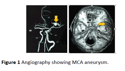
Figure 1: Angiography showing MCA aneurysm.
Aneurysms of middle cerebral artery represent almost a third of all the aneurysms of the circle of Willis anterior sector [1]. The lesions that cause aphasia are usually located in the left cerebral hemisphere. This is because the left hemisphere tends to be dominant for language, in both right handed and left-handed people [2]. Ochfeld et al. [3] showed that acute aphasia syndrome may allow the clinician to predict the compromised vascular territory, even when structural imaging shows only a small (or no) infarct. Even clipping is the ideal and only complete treatment of the intracranial aneurysm, wrapping is a well- known alternative technique when aneurysm clipping is not feasible, or is not completely satisfactory.
The Teflon material has been widely used in neurosurgical microvascular decompression and in cardiovascular surgery. In these fields, its reliability, safety, and lack of harmful effects have been widely recognised and should also apply in aneurysmal surgery [4].
21134
References
- Marques-Sanches P, Spagnuolo E, Martinez F, Pereda P, Tarigo A, et al. (2010) Aneurysms of the middle cerebral artery proximal segment (M1). Anatomical and therapeutic considerations. Revision of a series. Analysis of a series of the pre-bifurcation segment aneurysms. Asian J Neurosurg 5: 57-63.
- Ochfeld E, Newhart M, Molitoris BAJ (2010) Ischemia in Broca’s area is associated with Broca’s aphasia more reliably in acute than chronic stroke. Stroke 41: 325-330.
- Pelissou- Guyoyat l, Deruty R, Mottolesse C, Amat D (1994) The use of teflon as wrapping material in aneurysm surgery. Neurol Res 16: 224-227.

