Keywords
|
| Bilateral basal ganglia lesions; Diabetic uremic patients; Hypoglycemia; Neuroimaging findings |
Introduction
|
| Acute and subacute extrapyramidal movement disorders associated with uniform neuroimaging features of bilateral symmetrical basal ganglia changes are rarely reported in diabetic patients undergoing hemodialysis. Although metabolic and vascular factors are thought to play an important role in the pathophysiology, still remains unclear. Very few cases are reported with Wang et al. being the first to describe three patients in 1998 [1]. We would like to draw attention to this clinical syndrome with two cases of end stage renal disease (ESRD) showing common clinical and radiological features with the previously reported ones following episodes of severe hypoglycemia. |
|
Case 1
|
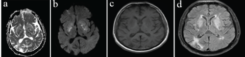
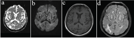
| A 63-year old male presented with sudden onset of hands tremor and gait imbalance. The patient had a 7-year history of type 2 diabetes mellitus, hypertension, ischemic stroke and ESRD with regular hemodialysis (thrice a week) since 16 months. He has been treated with insulin (NPH) 8 units and glimepiride 2 mg once daily in the morning. Neurological examination revealed dysarthria, dysphonia and quadriparesis. Low-amplitude bilateral upper limb resting tremor, bradykinesia and ataxic gait was present. Laboratory investigations indicated raised blood urea, 76 mg/dL (17-43) and serum creatinine, 8.3 mg/L (0.5-1.1) levels. Random blood glucose level on admission was 52 mg/ dl. The patient ingested a glucose tablet and blood glucose level measured the 20 min later was 98 mg/dL. The other findings were unremarkable. Serum parathormone, blood iron and blood lactate levels, and total iron binding capacity were also normal. Cranial magnetic resonance imaging (MRI) showed bilateral putaminal T1-hypointense and T2-hyperintense lesions and chronic ischemic areas at admission. The diffusion-weighted (DW) images revealed hyperintense/bright signal in the basal ganglia bilaterally with reduced apparent diffusion coefficient (ADC) (Figure 1a-d). During follow up the tremor and gait imbalance resolved within 7 days and recurrent episode of hypoglycemia was not observed. Follow-up cranial MRI performed 20 days after the first one, showed complete resolution of the bilateral basal ganglia lesions (Figure 2a-d). Endocrinologist was consulted and glimepiride was stopped due to the risk of inducing hypoglycemia, insulin injections were continued the patient’s blood glucose levels were well controlled. Regular thrice-weekly hemodialysis was maintained. Corticosteroids or any other medical treatment was not initiated. He remained free of tremor and gait imbalance during a 4-month follow-up. |
|
Case 2
|
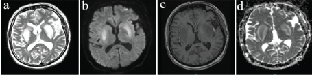
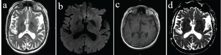
| A 46-year-old male patient presented with sudden onset of slurred speech, slow gait and loss of balance. His medical history revealed insulin resistance and ESRD. He was under regular hemodialysis (thrice a week) for 2 year and has been using metformin 1000 mg (1x1) since 6 months. Neurological examination revealed dysarthria, quadriparesis and left dorsal plantar response. Lowamplitude bilateral upper limb resting tremor prominent in the left side and postural instability was also present. Laboratory investigations indicated raised blood urea 126 mg/dL (17-43) and serum creatinine 8.72 mg/dL (0.5-1.1) levels. Random blood glucose level on admission was 47 mg/dL. IV %5 dextrose infusion was initiated and discontinued after the blood glucose level reached 98 mg/dl. The other findings were unremarkable. Serum parathormone, blood iron and blood lactate levels, and total iron binding capacity were normal. Cranial MRI showed bilateral T1- hypointense and T2-hyperintense putamen lesions at admission. The DW images revealed hyperintense/bright signal in the basal ganglia bilaterally with reduced ADC (Figure 3a-d). The use of oral antidiabetics was stopped and subcutaneous insulin treatment was initiated after consulting an endocrinologist. Corticosteroids or any other medical treatment was not initiated. The tremor and gait imbalance resolved within 7 days. After 25 days, follow-up brain MRI showed complete resolution of the lesions (Figure 4ad). Under regular thrice-weekly hemodialysis, he remained free of symptoms during a 7-month follow-up. |
Discussion
|
| Uremic patients can present with a wide variety of neurological complications which significantly contribute in morbidity and mortality. Uremic encephalopathy in patients with ESRD is a frequent and well-defined syndrome. Cerebral damage is prominent to the level of cortex, and patients often present with seizure and loss of memory and other cognitive abilities. |
| Involvement of the basal ganglia in uremia is rare with very few ESRD patients reported. The most common neurogical features include acute or subacute onset of parkinsonism and involuntary movements, mostly chorea. Mildly altered mental status, gait abnormalities, dysarthria, and difficulty in swallowing are less common [1,2]. |
| Neuroimaging features are the main characteristic of this condition. The vasogenic edema shown by DW MRI studies encounteres for these characteristic features. Our patients underwent detail MRI study using the DW images in addition to the standard T1- and T2-weighted images. Bilaterally hypointense lesions on T1-weighted and hyperintense lesions on T2-weighted series images on level of basal ganglia were detected. There was also evidence of central area cytotoxic edema surrounded by vasogenic edema in these affected regions. Similar observations were made by previously reported cases [3,4]. |
| The majority of patients with basal ganglia involvement are thought to be diabetic uremic patients and moreover in non-uremic diabetic patients the basal ganglia involvement is observed to be more frequent compared to non-diabetics. Whether it is uremic neurotoxines, metabolic acidosis or diabetic microangiopathy, the initiating event for the cytotoxic injury is still unknown. In 2010, Jury?czyk et al., suggested hypoglycemia as one of the candidate trigger factors causing this syndrome and emphasized the significance of strict glucose control in uremic patients [5]. Sangeon Gwoo et al also reported a similar case of acute bilateral basal ganglia lesions due to hypoglycemia in hemodialysis patient [6]. Our patients, one diabetic the other diagnosed with insulin resistance were both hypoglycemic on admission. |
| Selective involvement of the basal ganglia might be explained with the high metabolic requirements of the putamen and its sensitivities to metabolic changes. Significantly reduced metabolism of glucose in the bilateral basal ganglia, mainly in the putamen, was demonstrated in two cases by Wang et al., on 18F-fluorodeoxyglucose positron emission tomography (FDG-PET) examination [7]. It has been also assumed that these patients have already compromised cellular function due to long term diabetes and acute episodes of toxic or metabolic insult will lead to tissue damage and edema. Based on this assumption we believe that our patient’s episode of hypoglycemia might have triggered tissue edema. |
| The syndrome is usually self-limiting with spontaneous regression that is accompanied by clinical improvement. Although usually symptoms tend to spontaneously regress, it is believed that it might indicate poor prognosis and advanced disease [2,8]. In the study conducted by Wang and Cheng including cases with renal insufficiency, in the control images performed following the first month a regression of the lesions has been shown. Still clinically while residual symptoms were detected in 50% of patients, there has been no recovery in 20% of cases [9]. Also, similar to the majority of the reported cases, the basal ganglia lesions in our patients disappeared within 3 weeks to 2 months. Li et al. reported that there is no specific treatment for this condition thus we did not prescribed any treatment in addition to insulin injections and hemodialysis [10]. Still correction of uremia, metabolic acidosis and blood glucose levels along with supportive therapy has been the main focus of management. |
| We would like to highlight these two cases to raise awareness of this syndrome and to emphasize the importance of adequate dialysis, prevention and correction of hypotension, acid-base and electrolyte imbalances and hypo- and hyperglycemia factors. |
Figures at a glance
|
 |
 |
 |
 |
| Figure 1 |
Figure 2 |
Figure 3 |
Figure |
|

