Research Article - (2023) Volume 15, Issue 2
Probiotics in dermatological therapy and skincare
Dr. T. Vinotha*,
Nishat A and
Sakena Joseph
Microbiology, Hindusthan College of arts and science, Coimbatore, Tamilnadu, India
India
India
*Correspondence:
Dr. T. Vinotha, Microbiology, Hindusthan College of arts and science, Coimbatore, Tamilnadu,
India,
Tel: 9524911131,
Email:
Received: 30-Jan-2023, Manuscript No. ijddr-23-13431;
Editor assigned: 02-Feb-2023, Pre QC No. ijddr-23-13431;
Reviewed: 16-Feb-2023, QC No. ijddr-23-13431;
Revised: 18-Feb-2023, Manuscript No. ijddr-23-13431;
Published:
25-Feb-2023, DOI: 10.36648-0975-9344- 15.2-996
Introduction
Probiotics are characterized as “live microorganisms'' when
regulated insufficient sums, present a medical advantage on
the host”. The use of probiotics has shown promising effects
on the prevention and treatment of different inflammatory
skin disorders such as acne and atopic dermatitis. Although
probiotic bacteria have documented skin benefits, live cultures
are generally not preferred in cosmetics. Rather than including
live bacteria cultures, many of the probiotic skincare formulae
use bacteria fragments or metabolites. There is not currently
any science developed to support the idea that live cells are any
more effective when applied to the skin than these fragments. In
the near future, some brands using live bacteria might emerge.
We will briefly review the science supporting the use of topical
probiotics in dermatological therapy skincare products [1].
Nonetheless, such a large number of the items right now marked
as probiotics neglect to conform to the principal attributes.
In 1900 Louis Pasteur first identified the microorganisms
responsible for fermentation, finally bringing fitness to and
promoting the sturdiness of Bulgarian rural human beings in
the event that they consumed fermented ingredients which
included yoghurt and the bacteria they contained. He cautioned
that “lactobacilli may counteract the putrefactive outcomes of
gastrointestinal metabolism that contributed to infection and
getting older” whereas E.Metchnikoff associated the enhanced
longevity of Bulgarian rural people to the regular consumption
of fermented dairy products such as yoghurt. He suggested
that lactobacilli might counteract the putrefactive effects of
gastrointestinal metabolism that contributed to illness and
aging. Hippocrates declared, 2000 years earlier, that “death
sits in the bowels.” Metchnikoff considered the lactobacilli as
probiotics (“probios,”) conducive to life of the host as opposed to
antibiotics); probiotics could have a positive influence on health
and prevent aging [2]. During the neolithic period of the age of
the stone, the domestication of animals occurred and man began
to get fermented food. Probably serendipitous contaminations
in favorable environments played a major role. The corrective
business has expanded the quantity of items delegated probiotics.
The term prebiotics was introduced by Gibson and Rober Froidin
1995 to describe food supplements that are non-digestible by the
host but are able to exert beneficial effects by Selective stimulation of growth or activity of microorganisms that are present in the
intestine. The phrase probiotic (from the Latin pro and the greek
bio literally meaning “for existence”) was delivered by way of the
German scientist Werner Kolathin 1953 to designate “energetic
substances which are essential for a whole some development of
existence.” In 1965, this time period was utilized by Lily and Stilwell
in a special context to symbolize “materials, secreted with the aid
of one organism which stimulates the boom of any other.” More
particularly, Fulerin 1992 defined probiotics as ‘alive’ microbial
feed supplement which beneficially affects the host animal by
way of improving its intestinal microbial stability. While there are
a few expected applications for probiotics in private consideration
items, explicitly for oral, skin, and close consideration, legitimate
guideline of the naming and advertising norms is as yet expected
to ensure that shoppers are for sure buying a probiotic item. This
audit investigates the on-going business sector, administrative
angles and likely uses of probiotics in the individual consideration
industry [3].
Probiotics have various mechanisms of actional thought the
exact manner in which they exert their effects is still not fully
elucidated. These range from bacteriocin and short chain fatty
acid production, lowering of gut pH, and nutrient competition to
stimulation of mucosal barrier function and immune modulation.
The later in particular has been the subject of numerous studies
and there is considerable evidence that probiotics influence
several aspects of the acquired and innate immune response by inducing phagocytosis and IgA secretion, modifying T-cell
responses, enhancing Th1 responses, and attenuating the
responses. The skin is the largest organ in the human body. Its
primary function is to protect our bodies from external harm
by acting as a Physical barrier, with additional roles that include
regulation of the body [4].
Skin microbiome
Temperature, evaporation control, sensation, and storage of Lipids
and water, Skin continuously undergoes self-renewal, so resident
microbial cells are shed in the process. Most of the Microbes
found on the skin are commensal organisms and harmless to
healthy individuals; Infact, some are considered mutualistic.
Organisms confer health benefits to the skin by secreting
antibacterial substances, preventing pathogen colonization, and
influencing host immune responses. Recent researches related
to control of skin barrier functions prove close connection [5].
Physical, immunological, and cell biological characteristics of skin
and its bacterial population, Joshua Lederberg suggested using
the term “human microbiome” to describe the collective genome
of our indigenous Microorganisms (microflora) colonizing the
whole body in starting from this time, microbiologists and
dermatologists joined their efforts to identify and describe
different microorganisms colonizing human skin to estimate a
number of each population And to understand, which microbial
variety can cause one or another dermatological condition.
In general, the microbiome is defined as a collective genome
of microorganisms. Thus, the skin microbiome is a genome
of microorganisms present on skin, in which microorganisms
support complex relations. Most microorganisms found on
human skin are not dangerous for human health. Some of them
are even necessary for skin health. They release antibacterial
substances, prevent pathogenic colonization of skin and affect
its immunity [6]. According to held researches, three groups of
microorganisms colonizing human skin are found, they are as
follows:
Group.1 Transitional Microorganisms Regularly, Occasionally
appearing
Group.2 Temporal Microorganisms present on skin for a Short
period.
Group.3 Microorganisms permanently colonizing skin
There are various microflora i.e. bacteria and microscopic algae
and fungi, especially those living in a particular site or habitat.
The Skin Microflora in the context of bacteria, our skin can be
viewed as a cultural environment composition of which is mainly
a result of our genetics, diet, way of life and a region where
we live. Therefore, human skin is unique and accordingly, the
skin microbiome of each person is also unique [7-9]. The skin
microbiome is highly dependent on the micro environment
of the sampled site, a reflection on the physiology of skin.
Sebaceous sites such as the forehead have the lowest diversity,
and Propionibacterium species are the dominant organisms.
On the other hand, moist areas (e.g., armpits, navel, and groin)
constitute higher diversity of microbiota, with Staphylococcus
and Corynebacterium species as the predominant members [10- 15].Moreover, skin sites with greater bacterial diversity (e.g., Forearm, hand, and buttock) can harbour diversity as high as a
sort higher than that of the gut microbiome. The acidic condition
resulting from sebum degradation discourages pathogens from
invading and establishing in the skin. Personal hygiene is another
environmental factor that has a direct effect on the skin’s microbial
flora. Soaps, makeup, and skincare Products (e.g. Moisturizers)
alter skin conditions that in turn may influence the types of
microbes residing on the skin. On the contrary, in dry areas, skin
microbiomes include β-Proteobacteria and Flavobacteriales.
Various factors affect the microbial flora of the skin and they can
be generally categorized into host and environmental factors [16].
Skin barrier and microbiota act like a shield protecting the
organism against harmful effects of the outdoor environment.
There is well-balanced interaction between permanent and
temporary populations on skin. This balance continuously
depends on internal and external (including environmental)
factors that change composition of microbial populations on skin
and a function of the host skin barrier. Change of this balance is
characterized as dysbacteriosis occurrence of which can worsen
such chronic skin diseases as a topic dermatitis and psoriasis, or
acne. However, dysbacteriosis can be observed not only among
bacteria, imbalance between bacteria and commensal fungus
strain on head skin is observed in cases of patients disposed to
dandruff Cosmetics for skin: The increase in products termed
probiotic on the market does not necessarily equate With
areas onto celebrate the successful translation of science to
commerce and consumers. Too many products fail to comply
with the characteristics required to be called probiotic. Many
false claims and rampant misuse of the term has resulted in
mainstream consumer Channels providing incorrect information
to consumers. Probiotics are not inside us, not in Fermented
food, not necessarily better if there are more species or a higher
viable count. Formulations are being concocted not based on
research evidence but on marketing and what might appeal to
consumers [17-20].
Probiotics and skin microflora
The skin is able to act as a physical barrier exerting several
functions such as fluid homeostasis, thermoregulation, immune
responses, neurosensory functions, metabolic functions, and
primary protection against infection. The skin microflora plays
a significant role in competitive exclusion of pathogens that are
aggressive and provoke infection in the skin and in the processing
of skin proteins, free fatty acids (FFAs), and sebum. Interestingly
the resident microbiota may be regarded as “beneficial” to the
normal, healthy host, but may become dangerous to the host
with disturbed skin integrity. Microorganisms may have a role
even in a topic dermatitis (AD), eczema, rosacea, psoriasis, and
acne. Even if there are only very few studies pursuing a probiotic
approach for microflora-related skin disorders, it is intriguing to
suppose that topical probiotic application can be beneficial either
for preventing or for treating altered microflora-associated skin
diseases [21, 22].
Antimicrobial substances
The potential topical use of probiotic strains capable of producing
potent antimicrobials toxins (i.e.,bacteriocin, bacteriocin-like substances, organic acids, and H2O2) has received increasing
attention to successfully prevent pathogen adhesion and outcompete
undesired species (Ohetal.2006;Giloretal.2008).
Topical compositions containing probiotic bacteria, spores, and
extracellular product and uses thereof represented the basis of
the invention of Farmer (2005) suitable for topical application to
the skin which can be utilized to inhibit the growth of bacteria,
yeasts, fungi, viruses, and combinations of The invention also
Use of Probiotics for Dermal Application disclosed methods of
treatment and therapeutic systems for inhibiting the growth of
pathogens and combinations thereof, by topical application of
therapeutic compositions. The probiotic microorganisms may be
bacteria, yeast, or mold. Suitable probiotics should be selected
according to one or more particular properties, being the
preferred properties of their competitive exclusion of pathogenic
organisms from the surface to which they are applied, adherence
to human tissue, sensitivity to antibiotics, antimicrobial activity,
acid tolerance, and a high oxygen tolerance. In particular, the
method consists of different application modalities (i.e. Lotions,
spraying, wipe paper) of one or more probiotic microorganisms
to a wide variety of surfaces, such as human skin and hospital
equipment and fixtures, in an amount effective to, at least partly,
prevent their contamination, colonization, growth, and crosscontamination
by the pathogenic bacteria [23,24].
Materials and Methods
Isolation of Bacteria From
The medium which was selected for the lactic acid bacteria
was deMan, Rogosa, and Sharpe (MRS) agar medium. Rice was
cooked and excess water was drained. It was allowed to cool at
room temperature .It was soaked fully in water and stored in a
container. It was covered and left overnight at room temperature.
Fermented rice water was serially diluted and plated. Aloopful of
inoculums was taken from the plate and streaked on a MRS and
incubated at 37˚C for 24hrs. A loopful of samples was streaked on the sterile MRS agar Petri Plate by quadrant streaking method,
under aseptic conditions. After streaking all the Petri Plates,
they were incubated at 37°C for 24 to 48hr. After the incubation,
colonies were restreaked on the MRS agar Petri Plate for the
formation of isolated colonies. Then from these plates isolated
colonies were restreaked on MRS agar slants and stored at 4°C
[25] (Figure 1).
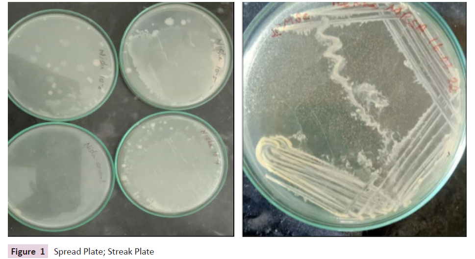
Figure 1: Spread Plate; Streak Plate
Identification of Bacteria: Lactobacilli confirmation can be done
microscopically by performing gram staining. The colour, shape
and motility were also observed.
Screening of Isolated Lactobacillus Spp. for
Probiotic Properties
Determination of optimal growth and pH: For the determination
of optimal pH and growth, the overnight culture of the Lactobacillus
in the MRS broth media with a varying pH ranging from 2.5 to 8.5,
using HCl or NaOH were inoculated with 1%(v/v) and incubated
in anaerobic condition for 24hr at 37°C in the presence of 10%
CO2. Bacterial growth was monitored by the determination of
optical density at 560 nm using a spectrophotometer against the
inoculated broth (Tables 1 and 2).
| Basic Characteristics |
Properties (Lactobacilusspp.) |
| Capsule |
Negative(-ve) |
| Catalase |
Negative(-ve) |
| Citrate |
Negative(-ve) |
| MR |
Negative(-ve) |
| VP |
Negative(-ve) |
| TSI |
Red Slant, Yellow butt, No Gas, No H2S |
| Gram staining |
Gram positive |
| Gas Production |
Negative(-ve) |
| Indole |
Negative(-ve) |
| Motility |
Mostly Negative(-ve) |
| Oxidase |
Negative(-ve) |
Table 1. Biochemical tests of Lactobacillus spp.
pH Value |
Sample (OD 560 nm) |
| 2.5 |
0.212 |
| 3.5 |
0.268 |
| 4.5 |
0.642 |
| 5.5 |
1.045 |
| 6.5 |
1.014 |
| 7.5 |
1.011 |
| 8.5 |
0.246 |
Table 2. Determination of optimal growth and pH.
Assay for NaCl tolerance: In order to determine the NaCl
tolerance, isolates from each sample were incubated with
1 % (v/v) overnight culture of Lactobacillus in the test tubes
containing MRS broth adjusted with different concentrations (1-
10%) of NaCl. The inoculated medium was incubated in anaerobic
condition for 24hr at 37°C. The bacterial growth was determined
using a spectrophotometer reading the optimal density at 560
nm (Table 3).
NaCl Concentration |
Sample (OD 560) |
| 1% |
1.258 |
| 2% |
1.183 |
| 3% |
1.096 |
| 4% |
1.074 |
| 5% |
0.948 |
| 6% |
0.917 |
| 8.5 |
0.346 |
Table 3. Assay for NaCl Tolerance.
Determination of bile salt tolerance: Bile salt tolerance of the
isolated bacteria was examined by inoculating the freshly cultured
isolates at1% (v/v) into MRS broth containing Bile salts different
concentrations (0, 0.1, 0.3, 0.5and1 %( w/v). The medium was
then incubated at 37°C for 24 hours in an anaerobic condition.
The growth of the isolates was monitored at 0, 3, 5 and 24hr by
measuring the absorbance of the culture broth at 620 nm using
spectrophotometer (Table 4).
Time in Hour |
0.10% |
0.30% |
0.50% |
1% |
| 0 |
0.218 |
0.218 |
0.219 |
0.21 |
| 3 |
0.211 |
0.261 |
0.203 |
0.126 |
| 5 |
0.374 |
0.354 |
0.116 |
0.152 |
| 24 |
0.923 |
0.571 |
0.096 |
0.086 |
Table 4. Determination of Bile Salt Tolerance.
Antimicrobial activity: The antimicrobial activity of the isolated
Lactobacillus was identified by well diffusion method, with
slight modification. The overnight culture of Lactobacillus was
centrifuged (10,000rpm for 20 min at 4°C) and the supernatant
was adjusted to pH7.0 with NaOH to exclude the antimicrobial
effect of H+. Supernatant of each sample of Lactobacillus were
monitored for antibacterial activity against indicator bacteria
(Bacillus, Pseudomonas) inoculated on Nutrient agar. Wells of
5mm India meter were punctured into the agar plate and aliquots
of 50μl from each solution samples were placed into the wells.
The plates were incubated for bacterial growth and examined for
the presence of clear zones of inhibition around the wells (Figure
2).
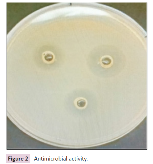
Figure 2: Antimicrobial activity.
Antibiotics sensitivity: For the observation of antibiotics
sensitivity test Disk diffusion method was performed. Different
antibiotics were tested over the isolates. The appropriate
antimicrobial-impregnated disks were placed on the surface of the MH agar plate inoculated with each isolate. The zones of
inhibition around the disks were observed and the diameters
were measured (Figure 3).
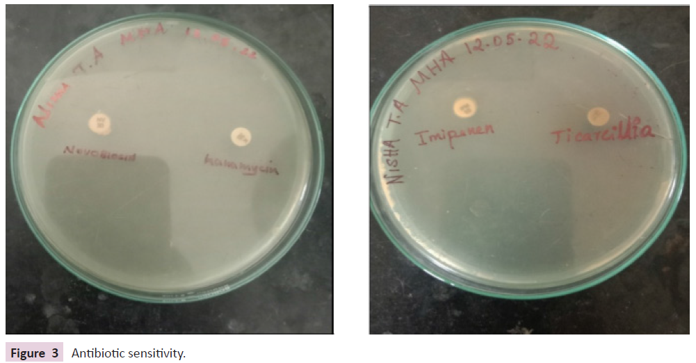
Figure 3: Antibiotic sensitivity.
Synthesis of probiotic bacteria: Isolated lactobacilli species was
in addition inoculated in nutrient agar broth. Inoculated broth
was incubated for 48hrs. For acquiring better concentration of
lactobacillus species, the broth became then centrifuged at 5000
rpm for 10min for pelleting down the classy cells. The pellet
became washed three times with autoclaved distilled water 3
instances after which it was weighed for determining the quantity
of mobile way of life received after incubation [26-28] (Figure 4).
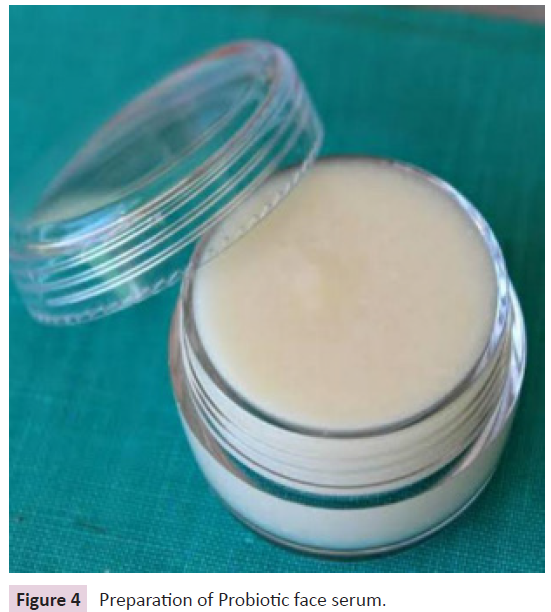
Figure 4: Preparation of Probiotic face serum.
Inoculation of Lactobacillus spp in Skincare
Shea butter is melted to liquid form. By double boiler over low
heat on the stove top, once it liquefies, heat is removed and
allowed to cool for 2 minutes then stir in the raw honey and aloe
vera gel, the mixture is allowed to cool off in the refrigerator for
about 15 minutes. Pellets of Lactobacillus species were added
and tea tree oil and essential oils were added drop by drop.
this probiotic face serum is transferred in sterilized glass jar and
stored in cool dry place The lab made face serum was further
dissolved in sterilized distilled water and a loopful was inoculated
to the Nutrient medium containing petri plates for determining
the viability of lactobacillus species after incubation [29] (Figure
5 and Table 1).
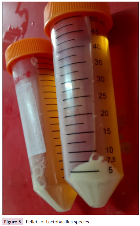
Figure 5: Pellets of Lactobacillus species.
Result and Discussion
MRS Medium plates were observed to have isolated bacterial
sample from fermented rice water. The bacterial sample was
further tested for confirmation of lactobacillus species. The
bacterial isolates were observed to be round, flat, slightly mucoid,
creamy in colour. Gram staining was performed and observed
to be gram positive bacteria and observed to be rod shaped.
Biochemical test was also performed [30] (Figure 6).
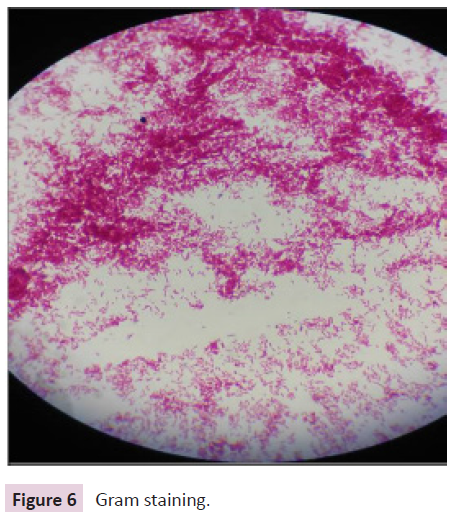
Figure 6: Gram staining.
Conclusion
This probiotic face serum is further tested to reduce inflammation,
Acne, And Atopic dermatitis People with atopic dermatitis
(AD), or atopic eczema, have a higher amount of the bacteria
Staphylococcus aureus in their skin the probiotic Lactobacillus
Spp. in the skin works on it. Analysis of the data indicated that it
reduced the number of S. aureus and decreased AD symptoms.
If it has anti acne and antimicrobial properties It prevents skin
from u radiation lactic acid is also Been described as Exfoliating
and moisturising agent probiotics will continue to expand in
applications and market share, and this exponential growth is
not likely to subside. It has been observed that existing cosmetic
products and their ingredients make use of viable cultures of
probiotics and/or their lysates for their production to maintain
good skin health. Their incorporation in certain cosmetics has
been proposed in order to achieve more radiant looking skin.
References
- Gasbarrini G, Bonvicini F, Gramenzi A (2017) Probiotics history. J Clin Gastroenterol 50:116-119.
Indexed at, Google Scholar, Crossref
- Van den Else LW, Poyntz HC, Weyrich LS, Young W, Forbes-Blom EE, et al. (2017) Embracing the gut microbiota: the new frontier for inflammatory and infectious diseases. Clin Trans Immunol 6(1):125.
Indexed at, Google Scholar, Crossref
- Salem I, Ramser A, Isham N, Ghannoum MA (2018) the gut microbiome as a major regulator of the gut-skin axis. Front Microbiol 9:14-59.
Indexed at, Google Scholar, Crossref
- Lee GR, Maarouf M, Hendricks AJ, Lee DE, Shi VY (2019) Topical probiotics: the unknowns behind their rising popularity. Dermatol Online J 25(5):13-30.
Indexed at, Google Scholar
- Tan AU, Schlosser BJ, Paller AS (2017) A review of diagnosis and treatment of acne in adult female patients. Int J Womens Dermatol 4(2):56-71.
Indexed at, Google Scholar, Crossref
- Platsidaki E, Dessinioti C (2018) Recent advances in understanding Propionic bacterium aces (Cutibacterium acnes) in acne. F1000Res 7:1000.
Indexed at, Google Scholar, Crossref
- Park SY, Kim HS, Lee SH, Kim S (2020) Characterization and analysis of the skin microbiota in acne: impact of systemic antibiotics. J Clin Med 9(1):168.
Indexed at, Google Scholar, Crossref
- Lee YB, Byun EJ, Kim HS (2019) Potential role of the microbiome in acne: a comprehensive review. J Clin Med 8(7):987.
Indexed at, Google Scholar, Crossref
- Peyri J (1912) Topical bacteria therapy of the skin. J Cutaneous Dis 30:688-689.
Indexed at, Google Scholar, Crossref
- Hendricks AJ, Mills BW, Shi VY (2019) Skin bacterial transplant in atopic dermatitis: known, unknowns and emerging trends. J Dermatol Sci 95(2):56-61.
Indexed at, Google Scholar, Crossref
- Di Marzio L, Cinque B, De Simone C, Cifone MG (1999) Effect of the lactic acid bacterium Streptococcus thermophilus on ceramide levels in human keratinocytes in vitro and stratum corneum in vivo. J Investig Dermatol 113:98-106.
Indexed at, Google Scholar, Crossref
- Bowe WP, Logan AC (2011) Acne vulgaris, probiotics and the gut- brain-skin axis-back to the future. Gut Pathog 3(1):1.
Indexed at, Google Scholar, Crossref
- Pavicic T, Wollenweber U, Farwick M, Korting HC (2007) Anti-microbial and -inflammatory activity and efficacy of phytosphingosine: an in vitro and in vivo study addressing acne vulgaris. Int J Cosmet Sci 29:181-190.
Indexed at, Google Scholar, Crossref
- Al-Ghazzewi FH, Tester RF (2010) Effect of konjac glucomannan hydrolysates and probiotics on the growth of the skin bacterium Propionic bacterium acnes in vitro. Int J/Cosmet Sci 32(2):139-142.
Indexed at, Google Scholar, Crossref
- Kang BS, Seo JG, Lee GS, Kim JH, Kim SY, et al. (2009) Antimicrobial activity of enterocins from Enterococcus faecalis SL-5 against Propionic bacterium aces, the Causative agent in acne vulgaris, and its therapeutic effect. J Microbial 47:101-109.
Indexed at, Google Scholar, Crossref
- Bowe WP, Filip JC, DiRienzo JM, Volgina A, Margolis DJ (2006) Inhibition of Propionic bacterium acnes by bacteriocin-like inhibitory substances (BLIS) produced by Streptococcus salivarius. J Drugs Dermatol 5:868-870.
Indexed at, Google Scholar
- Cosseau C, Devine DA, Dullaghan E, Gardy JL, Chikatamarla A, et al (2008) The commensal Streptococcus salivarius K12 downregulates the innate immune responses of human epithelial cells and promotes host-microbe homeostasis. Infect Immun 76:4163-4175.
Indexed at, Google Scholar, Crossref
- Lopes EG, Moreira DA, Gullón P, Gullón B, Cardelle-Cobas A, et al. (2017) Topical application of probiotics in skin: adhesion, antimicrobial and antibiofilm in vitro assays. J Appl Microbiol 122(2):450-461.
Indexed at, Google Scholar, Crossref
- Catherine Mack Correa M, Nebus J (2012) Management of patients with atopic dermatitis: the role of emollient therapy. Dermatol Res Pract 2012:836-931.
Indexed at, Google Scholar, Crossref
- Di Marzio L, Centi C, Cinque B (2003) Effect of the lactic acid bacterium Streptococcus thermophiles on stratum corneum ceramide levels and signs and symptoms of atopic dermatitis patients. Exp Dermatol 12:615-620.
Indexed at, Google Scholar, Crossref
- Gibson GR (1998) An overview of probiotics, prebiotics and symbiotic in the functional food concept: perspectives and future strategies. International Dairy Journal 8(5-6): 473-479.
Indexed at, Google Scholar, Crossref
- Toma MM, Pokrotnieks J (2006) Probiotics as functional food: microbiological and medical aspects. Acta Universitatis 710:117-129.
Indexed at, Crossref
- Salminen SJ, Gueimonde M, Isolauri E (2005) Probiotics that modify disease risk. Journal of Nutrition. 135(5):1294-1298.
Indexed at, Google Scholar, Crossref
- Fuller R (1999) Probiotics for farm animals Critical Review. Horizon Scientific 2:15-22.
Indexed at, Google Scholar, Crossref
- Fuller R (1989) Probiotics in man and animals. Journal of Applied Bacteriology 66(5):365-378.
Indexed at, Google Scholar
- Bockmuhl D (2006) Laundry and textile hygiene in healthcare and beyond. IFSCC 6:9:1-5.
Indexed at, Google Scholar, Crossref
- Marzio (2008) Increase of skin ceramide lev els in ac subjects. Int Jr Immunopathology Pharmacol 21(1):137-143.
Indexed at, Google Scholar, Crossref
- Zeeuwen PL (2013) Microbiome and skin diseases. Curr Opin allergy Clin immune 13:420-514.
Indexed at, Google Scholar, Crossref
- Holland KR (2002) Cosmetics- what is there influence on skin microflora? Am Jr Clin Derm 3(7):445-449.
Indexed at, Google Scholar, Crossref
- Salva A (2014) Role of the skin microbiome in atopic dermatitis. Clin Transl Allergy 4:33.
Indexed at, Google Scholar, Crossref
Citation: Vinotha T, Nishat A, Joseph S
(2023) Probiotics in Dermatological Therapy
and Skincare. Int J Drug Dev Res J, Vol. 15
No. 2: 996.


