Elichilia R Shao1, 2, 4*, Stanley G Ndazana2,Wilfred Chacha1,Gileard Masenga3,Sonda Tolbert1, 2,Dominic Mosha2, 5,Emmanuel G Kifaro1,2Balthazar M Nyombi1, 2
Kilimanjaro Christian Medical Center, PO BOX 3010 Moshi, Tanzania
Kilimanjaro Christian Medical University College, PO BOX 2240 Moshi, Tanzania
Kilimanjaro Christian Medical Center Gaenacology Outpatients, PO BOX 301Moshi Tanzania
Kiliamnjaro Christian Medical Center Molecular Diagnostic Unit, PO BOX 3010 Moshi Tanzania
Ifakara Health Institute, PO BOX 78373 Mikocheni, Dar es Salaam Tanzania
*Corresponding Author:
Elichilia R Shao
Kilimanjaro Christian Medical Center
PO BOX 3010 Moshi
Tanzania
Tel: +255784491622
E-mail: Elichilia2004@yahoo.co.uk
Keywords
Toxoplasmosis; Trimester; Congenital; Hydrocephalus; Northern Tanzania
Background
Toxoplasmosis is prevalent worldwide where by more than 70% of global population is infected with the parasite [1-4]. Human infection can result from ingesting undercooked meat containing Toxoplasma cysts. Another mode of transmission is from direct contact with cats’ litter or consuming food contaminated by oocysts excreted in the feces of infected cats. Its spread is influenced by various factors such as climate conditions, immune status and cultural behaviours of individual’s hygiene, type of food, cooking methods among others [4-6]. Presence of this infection during pregnancy, especially during the third trimester can lead to serious neonatal complications. The most pronounced complications are miscarriage, chorioretinitis, hydrocephalus, cerebral calcification and fetal death [7,8]. There is increased risk of congenital transmission among pregnant women in the third trimester as compared to first trimester [9,10]. There is uneven distribution of T. gondii prevalence among pregnancy and childbearing age from different parts of the world. In Europe, the prevalence of Toxoplasmosis varies between 20%-50% in the South and between 50%-70% in the West, whereas in the humid region of Africa, the prevalence varies [11]. The poor socioeconomic status in many countries in Latin America, Asia and Africa has been the primary risk factor as it has been association with poor hygiene and unreliable safe water use [12]. Presence of cat in the house and poor hygiene during handling of cats’ litter exposes these individuals to Toxoplasmosis infection especially for pregnant women who are the group of interest [10,12,13].
In Tanzania, only two studies have been conducted on the prevalence of Toxoplasmosis, in Dar es Salaam [14,15] the prevalence of Toxoplasmosis among pregnant women was 35% while Mwanza showed the prevalence of 30.9% respectively which seems to concur with other part of the world. This means more fetus are at increased risks to develop complications from Toxoplasmosis [16,17]. To prevent maternal-fetal transmission of Toxoplasmosis can be achieved through screening at the antenatal clinics and treat by using drugs such as spiramycine to prevents its complications [17]. Since there is no information on the prevalence of Toxoplasmosis among pregnant women in Kilimanjaro, this study was conducted in order to determine seroprevalence of T. gondii and associated factors to justify the need for screening among pregnant women in Tanzania.
Material and Methods
Study design and population
A cross-sectional hospital based study was conducted from April 26th to May 18th 2014 in Kilimanjaro Christian Medical Centre (KCMC) in North-eastern Tanzania. The hospital’s where this study was conducted; deliver services to about 15 million people in Northern part of Tanzania. In this region most of employed/business people consume pork and beef, this might be undercooked especially when grilled at bars. Permission to conduct this study was granted by Kilimanjaro Christian Medical University College ethical review committee. All women signed an informed consent prior to enrolment. Pregnant women attending antenatal care clinic were enrolled consecutively into the study during the study period. Inclusions criteria were all pregnant women who were residing in Moshi during study period while exclusion criteria were those who were residing in Moshi for less than three months. A full medical history including sociodemographic characteristics, presences of chronic illness and eating habit was recorded.
Samples
The samples were collected by aseptic procedures from each among the 144 pregnant women. About 4mls of venous blood were drawn in plain tubes, and transported from antenatal clinic to clinical laboratory. All samples were left at to coagulate for 30 minutes, and then separated by centrifugation at 3000 rpm for 4minutes to obtain sera. Sera were stored at -20 ºC till analysis.
ELISA reagents and procedure
All samples were tested for detection of IgG antibodies to T. gondii using ELISA commercial kit (Human Gesellschaft fur Biochemica und Diagnostica mbH, Max-Planck-Ring, Wiesbaden- German). Serum samples was diluted 1:100 (5μL: 450μL) with dilution buffer, 100μL of diluted samples were then applied to a coated microtiter plate, and incubated for 30 minutes at room temperature. The plate was then washed 4 times, with 350μ of 1:20 dilution washing buffer using automated ELISA washer (Lx50 BioTek, Highland park, Winooski, USA). Then, 100μL of conjugate was added, and incubated for 30 minutes, at room temperature, then washed 5 times as described above. 100μL of substrate solution was added, and incubated in dark for 15 minutes, and then the reaction was stopped by 100μL of stop solution. Plate was read at 450 nm soon after adding stop solution using ELISA reader (Lx800 BioTek, Highland Park, and Winooski, USA). Results were interpreted as positive when the sample OD ≥ MCC + 15%, and negative when sample OD < MCC – 15%, as per manufacturer instructions.
Rapid test
All positive samples from IgG ELISA assay were re-tested by using a rapid test kit (Toxo IgM/IgG lateral flow chromatography immunoassay, Houston, TX USA), a lateral flow chromatography assay which detect and differentiate between IgM and IgG antibodies to T. gondii in human serum, according to manufacturer’s instructions.
Data analysis
All statistical analyses were performed using a commercially available statistical package STATA version 12.1(StataCorp LP, College Station, TX). Categorical variables were summarized using cross tabulation to estimate different proportion. The effect of social-demographic characteristics and eating habit of meat on primary outcome of the study (Toxoplasmosis infection) was assessed using bivariate analysis. Explanatory variables were included in the multivariate analysis if the variable had p-value < 0.2 in bivariate analysis. Logistic regression model were used to estimate the odds ratio (OR) and 95% confidence interval (CI) to evaluate the strength of association between explanatory variable and primary outcome. Two sided Wald test p-value are presented.
Results
A total of 144 pregnant women were enrolled during the study period majority of them (55%) were aged 30yrs and above. About fifty six percent of the studied participants were from urban residence 81/144, only 36% were employed the rest were doing business and agriculture activities. On gravidity, about 87/144 (60.42%) were multigravid while 65/144(45.14%) were at their third trimester. Most of the study participants 82/144 (60%) had secondary education and above (Table 1). Of the 144 pregnant women studied, 60 (41.67%) were found seropositive for anti- Toxoplasma IgG antibodies, indicating chronic infection (Table 1). Out of seropositive study participants, one woman (0.69%) had anti-Toxoplasma IgM, indicating acute infection for Toxoplasmosis (Table 1).
| Patient’s Characteristic |
ELISA-IgG Test for T. gondii |
| Age group(years) |
Negative |
Positive |
| |
No |
Row% |
No |
Row% |
| 18-(n=65) |
31 |
47.7 |
34 |
52.3 |
| 30-(n=79) |
53 |
67.1 |
26 |
32.9 |
| Residence |
| Rural (n=63) |
37 |
58.7 |
26 |
41.3 |
| Urban (n=81) |
47 |
58.0 |
34 |
42.0 |
| Education |
| Primary level (n=62) |
30 |
48.4 |
32 |
51.6 |
| Secondary and above (n=82) |
54 |
65.9 |
28 |
34.1 |
| Occupation |
| Business (n=32) |
18 |
56.2 |
14 |
43.8 |
| Employed (n=52) |
36 |
69.2 |
16 |
30.8 |
| Peasant (n=60) |
30 |
50.0 |
30 |
50.0 |
| Gravidity |
| Primigravid (n=46) |
25 |
54.3 |
21 |
45.7 |
| Multigravid (n=87) |
52 |
59.8 |
35 |
40.2 |
| Grandmultigravid(n=11) |
7 |
63.6 |
4 |
36.4 |
| HIV status |
| Negative (n=125) |
73 |
58.4 |
52 |
41.6 |
| Positive (n=19) |
11 |
57.9 |
8 |
42.1 |
| Undercooked meat |
| No (n=99) |
57 |
57.6 |
42 |
42.4 |
| Yes(n=45) |
27 |
60.0 |
18 |
40.0 |
| Trimester |
| 1st Trimester (n=22) |
14 |
63.6 |
8 |
36.4 |
| 2nd Trimester(n=57) |
39 |
68.4 |
18 |
31.6 |
| 3rd Trimester (n=65) |
31 |
47.7 |
34 |
52.3 |
| Contact with cat |
| No (n=95) |
55 |
57.9 |
40 |
42.1 |
| Yes (n=49) |
29 |
59.2 |
20 |
40.8 |
Table 1: Distribution of T. gondii sero-prevalence with demographic characteristics among pregnant women (N=144), 2014 in Moshi.
The risks of contracting T. gondii was less by nearly 50% among pregnant women 30years and above compared with those below 30yrs (Crude OR=0.45: 95% CI 0.23-088; p=0.2). There was no statistical evidence for the difference (p=0.059) among T. gondii positive women across first and third trimester among studied pregnant women because the exactly time of infection was not defined (Figure 1). HIV infection was not statistically significant associated with T. gondii infection (adjusted OR 1.17: CI 0.42- 3.24; p=0.764 (Table 2). On multivariate logistic regression analysis there was significant difference in Toxoplasma seropositivity among pregnant women who are younger as compared to elderly ones (53.31% versus 32.91%) (OR=0.32, 95% CI: 0.23- 0.88, p=0.018). Potential factor HIV seropositivity was subjected into multivariate logistic regression analysis but there was no association. Occupation of the pregnant women was subjected to logistic regression analysis but there was no association even when was associated with residence, as majority of employed and business women were living in urban areas as compared to peasant who were residing in rural settings. Contact with domestic cat was exposed to both Univariate and multivariate logistic regression but there was no significant association with Toxoplasma infection among pregnant women.
| Variable |
Toxoplasmosis infection |
Crude OR
[95% CI] |
p-value |
Adjusted ORα
[95% CI] |
p-value |
| |
No
n (%) |
Yes
n (%) |
|
|
|
|
| Age (years) |
| Below 30 |
31 (36.9) |
34 (56.7) |
|
|
|
|
| ≥30 |
53 (63.1) |
26 (43.3) |
0.45 [0.23–0.88] |
0.020 |
0.41 [0.20–0.84] |
0.014 |
| Residence |
| Rural |
37 (44.0) |
26 (43.3) |
|
|
|
|
| Urban |
47 (56.0) |
34 (56.7) |
1.03 [0.53–2.01] |
0.932 |
1.18 [0.54–2.60] |
0.675 |
| Education |
| Primary |
30 (35.7) |
32 (53.3) |
|
|
|
|
| Secondary |
54 (64.3) |
28 (46.7) |
0.49 [0.25–0.96] |
0.036 |
0.64 [0.24–1.68] |
0.362 |
| Occupation |
| Peasant |
30 (35.7) |
30 (50.0) |
|
|
|
|
| Employed |
36 (42.9) |
16 (26.7) |
0.44 [0.02–0.97] |
004 |
0.61 [0.20–1.86] |
0.389 |
| Business |
18 (21.4) |
14 (23.3) |
0.77 [0.33–1.84] |
0.57 |
1.10 [0.42–2.89] |
0.841 |
| Gravidity |
| Primigravid |
25 (29.8) |
21 (35.0) |
|
|
|
|
| Multigravid |
59 (70.2) |
39 (65.0) |
0.79 [0.39–1.60] |
0.507 |
1.26 [0.51–3.09] |
0.620 |
| Trimester |
| First |
14 (16.7) |
8 (13.3) |
|
|
|
|
| Second |
39 (46.4) |
18 (30.0) |
0.81 [0.29–2.27] |
0.685 |
0.881 [0.31–2.74] |
0.881 |
| Third |
31 (36.9) |
34 (56.7) |
1.92 [0.71–5.19] |
0.199 |
2.52 [0.85–7.51] |
0.097 |
| HIV |
| Negative |
73 (86.9) |
52 (86.7) |
|
|
|
|
| Positive |
11 (13.1) |
8 (13.3) |
1.02 [0.38–2.71] |
0.967 |
1.17 [0.42–3.24] |
0.764 |
| Undercooked meat |
| No |
57 (67.9) |
42 (70.0) |
|
|
|
|
| Yes |
27 (32.1) |
18 (30.0) |
0.90 [0.44–1.85] |
0.784 |
0.79 [0.37–1.70] |
0.553 |
| Contact with Cat |
| No |
55 (65.5) |
40 (66.7) |
|
|
|
|
| Yes |
29 (34.5) |
20 (33.3) |
0.95 [0.47–1.91] |
0.882 |
1.04 [0.50–2.18] |
0.919 |
Table 2: Factors associated with T. gondii sero-prevalence among 144 pregnant women 2014 in Moshi.
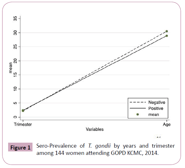
Figure 1: Sero-Prevalence of T. gondii by years and trimester among 144 women attending GOPD KCMC, 2014.
Discussion
This was among few studies in Tanzania, and the first study to be conducted among pregnant women in North-eastern part of the country to determine the actual scenario of Toxoplasmosis among this population. The seroprevalence of T. gondii infection in this study was found to be higher than lake zone (30.9%) and (35%) reported in Mwanza and Dar es Salaam respectively. But the prevalence was lower compare to 46% among abattoir workers, livestock keepers and animal health workers in Tanga. The different in prevalence might be explained by the fact that, these people were exposed to animal and soil environment hence more chance to contact infection as compared to pregnant women from different professionals [15]. The different could partly be explained by climatic conditions where Moshi has more rainy seasons compared with Dar es Salaam and Mwanza Tanzania. This conditions favours sporulation of oocysts; the methodology which was used in the current was ELISA which is more specific as compared to immunosorbent agglutination which was used in Dar es Salaam [16,17]. Age different among study participants was also associated with Toxoplasmosis infection; where by younger women were more infected compared to elderly ones. This finding was different from other studies which showed that there was an increased risk of infection as age increases [18]. This might be explained by the facts that younger people prefer outing compared to elderly, this outing expose them to grilled meat (which might be undercooked), fruits and saladies which may be contaminated with the parasites hence increased risks of infection [19-21]. In the current study we could not extrapolate exactly time of infection, across three trimester, which is one of the weakness of our study compared to other studies. [10,22]. Contact with cat litter is another contributing risk for T. gondii infection [23], in the current study about one third of the study participants were keeping house cat but there was no significant association between T. gondii infection and history of cat contact was found. This is because presence of cat in the house is not enough to confirm zoonosis, rather handling of cats’ litter is of more important. These findings were consistence with studies which was conducted in Mwanza Tanzania [17], Palestine [24] and Zambia [25]. But other studies from Ethiopia [26] and Taiwan [27] showed a significant association between contact with cats and seroprevalence of T. gondii. The presence of one woman with active infection who wasn’t aware with the disease means more risks to the fetus, this stress on the significant of screening to control congenital transmission and its outcome. Apart from consequences among pregnant women [28], in general population Toxoplasmosis is associated with risks to develop schizophrenia and reduce intelligence which compromise social adaption of infected person [4,29]. Immune compromised people are more prone to clinical Toxoplasma [30], in the current study about thirteen percent of the studied women were HIV positive but there was no statistical significant association between HIV seropositivity and positive ELISA for Toxoplasmosis. This might be partially be explained by small sample size as well as most of the women were on ART hence reduced chance for the opportunistic infections.
Conclusions
This study confirmed seroprevalence of T. gondii among pregnant women in North-eastern Tanzania, which suggests the need for intervention, especially with regards to education on the associated factors for the purpose of reducing morbidity and mortality. Despite the lack of statistical correlation between infection and associated risk factors, preventive measures among pregnant women should be followed to prevent consequences of Toxoplasmosis.
Author’s Contributions
ERS designed the project. ERS, EGK, GM and SNN collected specimens and clinical data. EGK and SNN analysed and interpreted the results. ERS, TS and DM analysed and interpreted the data. ERS wrote the draft of the manuscript which was approved by all authors. All authors read and approval the final version of this manuscript.
Acknowledgments
The authors would like to thank all staff in KCMC antenatal care clinic and Serology Unit for their technical support.
6690
References
- Cenci-Goga BT, Rossitto PV, Sechi P, McCrindle CM, Cullor JS (2011) Toxoplasma in animals, food, and humans: an old parasite of new concern. Foodborne Pathog Dis 8: 751-762.
- Halonen SK, Weiss LM (2013) Toxoplasmosis. Handb Clin Neurol114: 125-145.
- Doudou Y, Renaud P, Coralie L, Jacqueline F, Hypolite S, et al. (2014) Toxoplasmosis among pregnant women: High seroprevalence and risk factors in Kinshasa, Democratic Republic of Congo. Asian Pac J Trop Biomed 4: 69-74.
- Paul RT, Pierpaolo M (2013) The global burden of congenital Toxoplasmosis: A systematic review. Bull World Health Organization 91: 501-508.
- Caballero-Ortega H, Uribe-Salas FJ, Conde-Glez CJ,Cedillo-Pelaez C, Vargas-Villavicencio JA, et al. (2012) Seroprevalence and national distribution of human toxoplasmosis in Mexico: analysis of the 2000 and 2006 National Health Surveys. Trans R Soc Trop Med Hyg 106: 653-659.
- Dabritz HA, Conrad PA (2010) Cats and Toxoplasma: implications forpublic health. Zoonoses Public Health 57: 34-52.
- Walle F, Kebede N, Tsegaye A, Kassa T (2013) Seroprevalence andrisk factors for toxoplasmosis in HIV infected and non-infected individuals in Bahir Dar, Northwest Ethiopia. Parasit Vectors6: 15.
- Torgerson PR, Macpherson CN (2011) The socioeconomic burden of parasitic zoonoses: Global trends. Vet Parasitol 182:79-95.
- Endrias ZG, Anteneh H, Tesfaye S, Kassu D Girmay M, et al. (2013) Seroepidemiology of Toxoplasma gondii infection in women of child-bearing age in central Ethiopia. BMC Infect Dis 13.
- Bessières MH, Berrebi A, Cassaing S, Fillaux J, Cambus JP, et al. (2009) Diagnostic of congenital toxoplasmosis: prenatal and neonatal evaluation of methods used in Toulouse UniversityHospital and incidence of of congenital toxoplasmosis.MemInst Oswaldo Cruz 104: 389-392.
- Wang T, Liu M, Gao XJ, Zhao ZJ, Chen XG, et al. (2011) Toxoplasma gondii: the effects of infection at different stages of pregnancy on the offspring of mice. Exp Parasitol 127: 107-112.
- Mpiga MR, Akue JP, Biskigou U, Mayi TS, Nkoghe D (2010) Serological study in pregnant women of Franceville, Gabon. Bull Soc Pathol Exot 103: 41-43.
- Khurana S (2010) Serological screening for antenatal oxoplasma infection in India. Indian J. Med. Microbiol 28: 143-146.
- Jones JL (2009) Risk factors for Toxoplasma gondii infection in the United States. Clin Infect Dis 49:878-884.
- Liu Q (2009) Toxoplasma gondii infection in pregnant women in China. Trans. R. Soc. Trop. Med Hyg 103:162-166.
- Swai E (2009) Seroprevalence of Toxoplasma gondii infection amongst residents of Tanga District in north-east Tanzania. Tanzania Journal of Health Research 4.
- Doehring E, Reiter-Owona I, Bauer O, Kaisi M, Hlobil H, et al.(1995) Toxoplasma gondii antibodies in pregnant women and theirNew borns in Dar es Salaam, Tanzania. Am J Trop Med Hyg52:546-548.
- Berno M, Stephen E, Benson R, Anthony N, Humphrey D, et al. (2013) Sero-prevalence and factors associated withToxoplasma gondii infection among pregnant women attending antenatal care in Mwanza,Tanzania. BMC parasit vectors 6: 222.
- Makiko S,Shunichi N, Masachi H, Hirotoshi N, Satoshi H (2011) Anti-Toxoplasma Antibody Prevalence, Primary Infection Rate, and Risk Factors in a Study of Toxoplasmosis in 4,466 Pregnant Womenin Japan. J of clinical and vaccine immunology 365-367.
- Koskiniemi M, Lappalainen M, Hedman K (1989) Toxoplasmosis needs evaluation.An overview and proposals. Am J Dis Child143:724-728.
- Ghoneim NS, Hassanain N, Zeedan G, Soliman Y, Abdalhamed A (2009) Detection of genomic Toxoplasma gondii DNA and anti-Toxoplasma antibodies in high risk women and contact animals. Global Veterinaria 3:395-400.
- Sroka JW-FA, Szymanska J, Dutkiewicz J, Zajac V, Zwolinski J (2010) The occurrence of Toxoplasma gondii infection in people and animals from rural environment of Lublin region -estimate of potential role of water as a source of infection. Ann Agric Environ Med17:125-132.
- Dabritz HA, Conrad PA (2010) Cats and Toxoplasma: implications for public health. Zoonoses Public Health 57: 34-52.
- Wang T, Liu M, Gao XJ, Zhao ZJ, Chen XG, et al. (2011)Toxoplasma gondii: the effects of infection at different stages of pregnancyon the offspring of mice. Exp Parasitol 127: 107-112.
- Nijem KA-AS (2009) Seroprevalence and associated risk factors of toxoplasmosisin pregnant women in Hebron district, Palestine. East Mediterr Health J 15:1279-1284.
- Ishaku B, Ajogi I, Umoh J, Lawal I, Randawa A (2009) Seroprevalence and risk factors for Toxoplasma gondii infection among antenatal women in Zaria, Nigeria. Res J Medicine & Med Sci 4:483-488.
- Endalew Z, Delenasaw Y, Solomon A, Tariku B, Abdi S, et al. (2012) Seroprevalence of Toxoplasma gondii and associated risk factors among pregnant women inJimma town, Southwestern Ethiopia. BMC inf Dis 12: 337.
- Lin YL, Liao YS, Liao LR, Chen FN, Kuo HM, et al. (2008) Seroprevalence and sources of Toxoplasma infection among indigenous and immigrantpregnant women in Taiwan. Parasitol Res 103:67-74.
- Ayi I, Edu A, Apea-Kubi K, Boamah D, Bosompem K, et al. (2009) Seroepidemiology of toxoplasmosis amongst pregnant women in the greater Accra region of Ghana. Gh Med J43:107-114.
- Dogruman-Al F, Aslant S, Alcan S, Customer S, Turk S (2009) A possible relationship between Toxoplasma gondii and Schizophrenia: A seroprevalence study. Int J psychiatr Clin Pract 13: 82-87.


