Hadi Mutairi*
Thoracic Surgery, King Fahad Specialist Hospital, Dammam, Saudi Arabia
*Corresponding Author:
Hadi Mutairi
Thoracic Surgery
King Fahad Specialist Hospital
Dammam, Saudi Arabia
Tel: 00966556372222
E-mail: hadimutairi@mail.com
Received date: March 07, 2016 Accepted date: March 12, 2016 Published date: March 19, 2016
Clinical Description
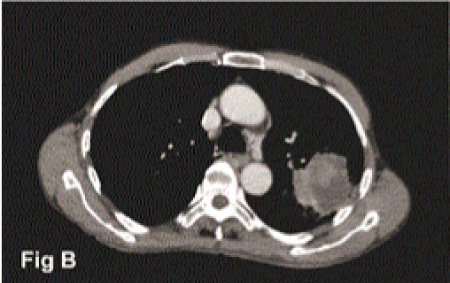
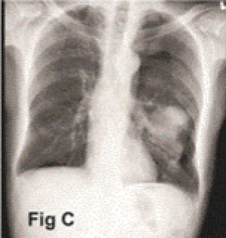
Clinical staging for lung cancer is a dynamic process. Information gained during the course of the work up of patients with lung cancer help the treating physician tailor the treatment plan accordingly. Lung cancer patient may present with a peripheral mass that may bring the question whether it invades the chest wall or not. This question has a significant impact on surgical treatment decision. Most of lung cancer patients nowadays undergo image-guided transthoracic needle biopsy to obtain a tissue diagnosis. Pneumothorax is a possible complication of such procedure. The incidence of such complication is rare and is related to many factors like needle size, location of the tumor, and the technique itself. We report a case of 45-year-old male, heavy smoker, with a left upper lobe peripheral lung cancer (Figures 1 and 2) in which a questionable chest wall involvement was refuted based on an incidental occurrence of pneumothorax after a needle biopsy (Figure 3). This is based on the fact that if the tumor is truly fixed to the chest wall, then pneumothorax would not have had happened after the biopsy. The biopsy result came back as primary non-small cell lung cancer, squamous type. The thoracic oncology tumor board decision was to proceed directly to surgical resection after completion of staging work up. The patient underwent a video assisted thoracoscopic (VATS) lobectomy and systematic mediastinal lymph node dissection, obviating the need for open procedure or the need for chest wall resection. There were no indications in this case to recommend adjuvant chemotherapy. At 2 year follow up, the patient showed no recurrence and is doing well.

Figure 1: A chest x-ray showing a tumor in the left upper lobe with possibility of lateral chest wall involvement.

Figure 2: A CT can cut showing the left upper lobe tumor with suspected chest wall invasion.

Figure 3: A chest x-ray after biopsy showing a left-sided pneumothorax with the tumor in the left upper lobe separated from chest wall indicating no invasion to the lateral chest wall.
This case is worth reporting because it highlighted the importance of paying attention to the details of every step in the process of staging of lung cancer. We do not know precisely how often does this happen but in our center this is the second incidence since we started our thoracic oncology program i.e., 7 years, with an average volume of 110 cases annually.
8776


