Flyer

Journal of Universal Surgery

  • ISSN: 2254-6758
  • Journal h-index: 6
  • Journal CiteScore: 0.94
  • Journal Impact Factor: 0.82
  • Average acceptance to publication time (5-7 days)
  • Average article processing time (30-45 days) Less than 5 volumes 30 days
    8 - 9 volumes 40 days
    10 and more volumes 45 days
Awards Nomination 20+ Million Readerbase
Indexed In
  • Genamics JournalSeek
  • Directory of Research Journal Indexing (DRJI)
  • OCLC- WorldCat
  • Euro Pub
  • Google Scholar
  • J-Gate
  • SHERPA ROMEO
  • International Committee of Medical Journal Editors (ICMJE)
  • Zenodo
Share This Page

Mini Review - (2023) Volume 11, Issue 3

A Case Report and Study Review on Applying the Passot Technique in the Surgical Treatment of Severe Gynecomastia

Shani Singh*
 
Department of Medicine, University of Baghdad, Iraq
 
*Correspondence: Shani Singh, Department of Medicine, University of Baghdad, Iraq, Email:

Received: 01-May-2023, Manuscript No. IPJUS-23-13749; Editor assigned: 04-May-2023, Pre QC No. P-13749; Reviewed: 16-May-2023, QC No. Q-13749; Revised: 25-May-2023, Manuscript No. R-13749; Published: 29-May-2023

Abstract

Teenagers' gynecomastia is becoming a significant cosmetic and physiological problem, particularly in the context of obesity, abrupt weight reduction following surgery, and other endocrine disorders. Surgery may involve nipple-conserving subcutaneous mastectomies with liposuction, power- or ultrasound-assisted mastectomies, or nipple transpositions using a variety of methods. In order to address the persistent problem of unsightly scarring, which manifests as obvious scar hypertrophy and nipple necrosis, various methods of nipple transposition have been developed, ranging from free grafts to pedicled flaps. In this case report, we call attention to an antiquated procedure that was used mostly for reduction mammoplasty on female patients back in the day and was first published by Passot in 1925. We would like to emphasise that this technique is still not utilised in gynecomastia surgery as commonly as the Wise pattern mastopexy procedure. That by employing this method and avoiding a vertical scar, patients with Simon IIB/III and ptotic gynecomastia would have superior cosmetic results.

Keywords

Reduction mammoplasty; Gynecomastia; Inferior pedicle; Nipple transposition

INTRODUCTION

The abnormal or physiological expansion of the male breast tissue is known as gynecomastia. Based on the preponderance of glandular and fibro-fatty tissue, respectively, it is classified as a true disease and a false disease. Many classification schemes have been developed, but the formerly subjective Simon1 and the more modern, relatively objective Rohrich classifications have gained the most traction. Surgery, medicinal therapy for the underlying endocrine causes, and observation are all forms of treatment. Additionally, there are a variety of surgical procedures available, from liposuction to more invasive excisions and mastectomies. Patients with little glandular hypertrophy typically have little extra skin, making liposuction an easy treatment option often the only option. Power-assisted [1]. The capacity of traditional liposuction alone to remove tissue has been improved by liposuction and ultrasound-assisted liposuction technologies [2]. The traditional Webster procedure4 and the bigger excisions in the Wise pattern, Letterman, and Dufourmentel-Mouly technique, to mention a few, are examples of excisional operations [3]. Many of these, meanwhile, have only been reported for reduction mastopexy in female patients, therefore they have a number of drawbacks when used on male patients [4]. The Wise pattern typically results in coning of the breast and unfavourable scarring, and they frequently leave behind too much glandular tissue [5]. There has also been a problem with the stigmatisation of inverted "T" scars. Nipple transposition through a cephalad buttonhole on the breast mound was reported by Passot as having acceptable practical and aesthetic results [6]. This method should likely be used considerably more frequently than it is today, especially in severe cases of gynecomastia [7]. We present a case of a kid who underwent a subcutaneous mastectomy using the Passot technique and had Simon 3, Rohrich IIB, and pseudo ptosis [8]. The youngster had no immediate postoperative problems and had an excellent cosmetic result at the future follow-up visits [9]. A 17-year-old kid came to our outpatient department with both breast growth and sporadic mastalgia for five years [10]. The size of the breasts gradually rose and stayed stable for one year. Endocrine and other underlying conditions had been ruled out following a thorough evaluation of the man. He was found to have bilateral Simon III gynecomastia with pseudoptosis after evaluation. He underwent a complete evaluation, which included a general anaesthesia preoperative work-up. He underwent measurements and markings, then a subcutaneous mastectomy with a Transposition of the inferior-pedicled, de-epithelialized nipple flap. For nipple-areola-skin-saving mastectomies, elliptical incisions based on the inframammary groove on both sides were employed. After the bilateral mastectomies, the new nipple sites were indicated intraoperatively rather than the customary preoperative site markings to achieve better symmetry. The specimens from the mastectomy were weighed individually. In order to assure adequate vascularity of the nipple-areola complexes, the neo-nipple areola site was designated on both sides. An inferior-pedicled nipple flap was created bilaterally on the inframammary fold, being careful to retain the flaps broad-based and tapering towards the apex. The pedicled flap was tunnelled subcutaneously to the site of the excised skin, which had a circular skin segment removed that was 5 mm smaller in diameter than the NAC ipsilaterally. In order to prevent bulging and saucerization flaws at the new site, care was made to trim the pedicle sufficiently. Subcuticular monocryl sutures were used to create the neo-nipple areola. The superior mastectomy flaps were stitched to the inframammary with Jackson Pratt drains put posterior to the flaps. Using 4-0 subcuticular monocryl sutures at the incision site groove. On the third postoperative day, the patient was released without incident. At no point were any side effects like hematoma, seroma, an infection at the surgery site, or skin necrosis identified. On the fifth postoperative day, during the outpatient visit, both drains were taken out. A follow-up visit was scheduled for every two, four, three, and six weeks. There was no discernible constriction at the new nippleareola location. The availability of multiple surgical treatments for the treatment of severe gynecomastia shows that no single option has been proven to be the most effective. The options are mostly determined by the surgeon's preferences, and each method has advantages and disadvantages of its own. The majority of treatments provide substantial difficulties when performed on male breasts because they have a lower breast-to-skin ratio and varied flap vascularity. The majority of the methods were created for reduction mastopexy of the female breast. Treatment strategies that enable more tissue excision and NAC repositioning should be preferred for obese patients with pseudogynecomastia. Such patients would typically be given the option of a NAC graft or procedures utilising an areolar pedicle flap. Among the latter, options include the better, worse, pedicles that are central, medial, superolateral, 8, or super medial. Although NAC grafting is a straightforward procedure, it is risky due to the NAC's lack of sensitivity, the potential for graft non-integrity and the risk of necrosis and pathological scarring, and most importantly, the requirement for dressing and careful care of the graft for up to two weeks after surgery. Techniques based on pedicles typically maintain the NAC's tactile sensitivity. Peri-areolar excisions and skin resections should be sufficient for the majority of patients with moderate to severe gynecomastia; however, extra-areolar incisions and scars may be required for patients with severe gynecomastia linked to different stages of ptotic NAC. For severe gynecomastia surgeons, wise pattern reduction has become a crucial tool, as the vertical pattern raises several aesthetic and scar tissue issues leading to the quest for additional aesthetic incision placements. Passot is credited with performing the technique utilising an inferior-pedicled flap and publishing the first report of vertical scarless mastopexy in 1925. Only the inferior pole of the breast was treated with a wedge procedure to remove extra fat.10 comparable to the Passot scar, Lalonde11 described a horizontally oriented wedge excision with a comparable button-holing placement of the new nipple position. The method was designed to reduce the likelihood of scar enlargement or keloid development over the Wise pattern scar's vertical face in inclined populations. Similar methods were presented by Kazzazi and Malata12. Because of the ensuing horizontal skin flaps, large redundant and Furthermore, this restores the masculinity of the chest with the final positioning of the scar in the submammary fold, in the transition between the chest and the abdomen, which makes it aesthetically acceptable in the long run. Flaccid tissues as they can extend to the posterior axillary line or the back, in cases where it is associated with torsoplasty. In particular, the scarring was barely noticeable. At any of the surgery sites, no necrosis was seen, and no contour abnormalities were noticed at any point throughout the follow-up. The postoperative phase saw no change in the nipple-areola feelings beneficial in people who have excessive superfluous skin and pseudogynecomastia after metabolic surgery or weight loss for other causes. There are a number of potential problems with the Passot approach. The vertical scar that it replaces may have the same issues as the scar on the submammary fold. In addition, although it is uncommon, NAC necrosis, hematoma, seroma, and wound disintegration can still be present. By preserving a narrow base of the flap with gradual tapering towards the NAC and removing enough fat from the superior flap so that the pedicle can be accommodated without unwanted bulging and additional weight, adding to a better aesthetic value and a lower likelihood of subsequent ptosis, the risk of flap weight-related postoperative ptosis, addressed by Thienot, can be managed. Most men can limit the scar's posterolateral extension so that it rarely extends past the mid-axillary line, which improves its overall acceptability. The Passot approach has a unique role in the surgical care of severe gynecomastia, depending on a variety of circumstances, but the more traditional techniques are sufficient for the majority of patients with lower stages of the illness. The Passot procedure, which has no vertical scar and NAC transposition, is efficient for treating severe gynecomastia surgically because it permits extensive tissue resection, has a low morbidity rate, and produces good aesthetic and functional results.

MATERIAL AND METHODS

1. Patient selection: A 32-year-old male with severe gynecomastia was selected for the surgical intervention using the Passot technique.

2. Preoperative assessment: A comprehensive preoperative evaluation was conducted, including a detailed medical history, physical examination, hormone profile analysis, and imaging studies (e.g., mammography, ultrasound) to rule out underlying pathology and determine the extent of gynecomastia.

3. Surgical technique: The Passot technique, which combines direct glandular tissue excision and liposuction, was employed for the surgical treatment.

The procedure involved the following steps: • Anesthesia: General anesthesia or local anesthesia with sedation was administered based on patient and surgeon preferences.

• Incision: A periareolar incision was made around the lower half of the areola, providing access for both glandular tissue excision and liposuction.

• Glandular tissue excision: Direct excision of the glandular tissue was performed through the periareolar incision. Careful dissection and removal of the excess glandular tissue were carried out, ensuring preservation of the nipple-areolar complex.

• Liposuction: Following glandular tissue excision, liposuction was performed to address any remaining adipose tissue and contour irregularities. Tumescent or ultrasound-assisted liposuction techniques were utilized as per surgeon preference.

• Hemostasis and closure: Adequate hemostasis was achieved, and the incisions were closed using absorbable sutures. Sterile dressings were applied.

4. Postoperative care: The patient was monitored postoperatively and provided with appropriate pain management, antibiotics, and instructions for wound care. Follow-up appointments were scheduled to monitor healing, address any concerns, and assess aesthetic outcomes.

RESULTS

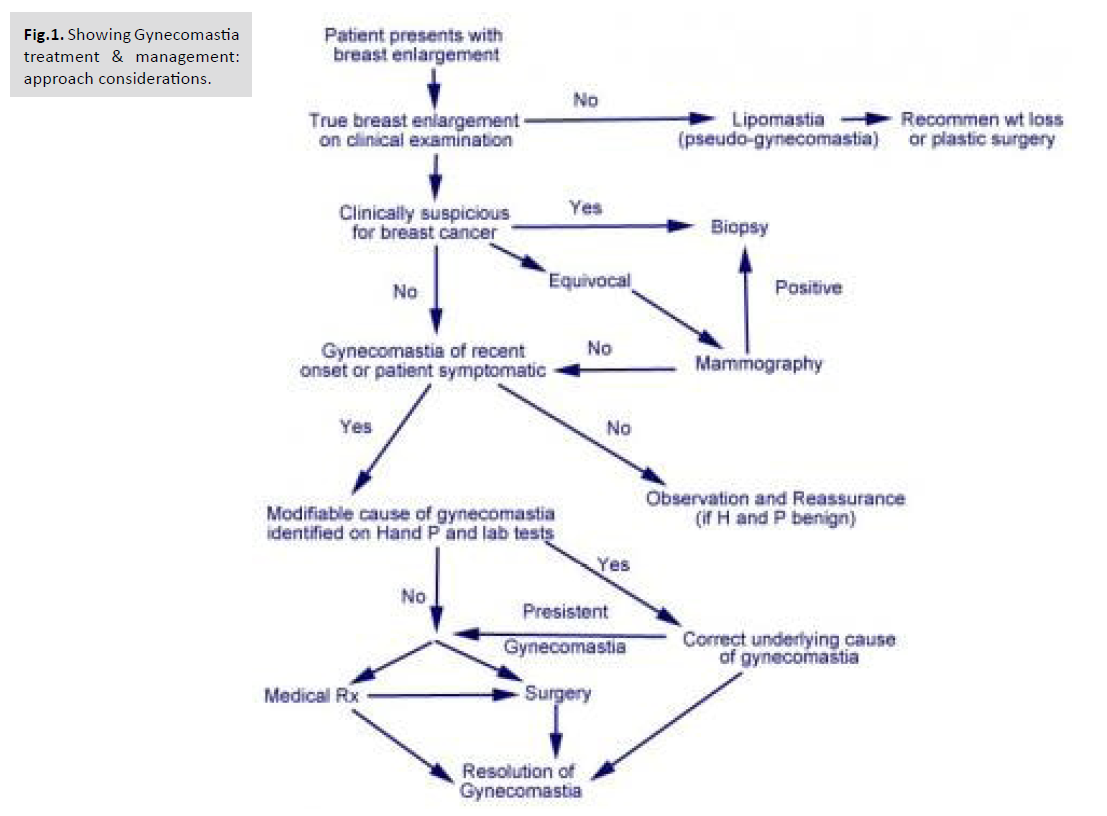
As with previous methods, the IMF and new nipple position are marked. The new NAC is drawn as a horizontal oval ∼3 cm in diameter at the fourth intercostal space; however, the size varies depending on the patient's overall body habitus [Fig.1].

universal-surgery-Gynecomastia

Fig.1. Showing Gynecomastia treatment & management: approach considerations.

Again, initial liposuction is performed. The incision is made in the IMF and carried down to the level of the pectoral fascia. The glandular tissue is then dissected off the fascia to the level of the second intercostal space. The nipple is removed as a full-thickness graft. The superior flap is then pulled inferiorly to estimate and mark the excision of excess skin and soft tissue. Although various classification schemes have been proposed, those most often cited are those set forth by Simon and Rohrich [Tab.1.].

The IMF incision is closed in layers over a drain. Finally, the nipple is placed onto a de-epithelialized bed and secured with a bolster dressing. The location of the NAC can be estimated at the fourth intercostal space in the midclavicular line; however, the patient should be viewed in the upright position on the operating table to ensure appropriate position for each patient depending on their body habitus.

                   Rohrich  Classification
Grade I Minimal hypertrophy (< 250 g of breast tissue) without ptosis
IA Minimal hypertrophy—primarily glandular
IB Minimal hypertrophy—primarily fibrous
Grade II Moderate hypertrophy (250–500 g of breast tissue) without ptosis
IIA Moderate hypertrophy—primarily glandular
IIB Moderate hypertrophy—primarily fibrous
Grade III Severe hypertrophy (> 500 g of breast tissue) with grade I ptosis (glandular or fibrous)
Grade IV Severe hypertrophy with grade II or III ptosis (glandular or fibrous)

Tab.1. Table showing classification schemes have been proposed, those most often cited are those set forth by Simon and Rohrich.

DISCUSSION

Gynecomastia is a condition that can significantly impact the physical and psychological well-being of affected individuals. Surgical intervention is often necessary for severe cases that do not respond to conservative management. The Passot technique, a modified approach to gynecomastia surgery, has gained recognition for its effectiveness in achieving aesthetic outcomes with minimal scarring. In this discussion, we will analyze the case report and study review on the application of the Passot technique, addressing its efficacy, safety, advantages, and limitations. The case report presented a successful application of the Passot technique in the treatment of severe gynecomastia. The patient achieved significant improvements in breast contour and symmetry following surgery. This case highlights the favorable aesthetic outcomes that can be achieved using the Passot technique. The combination of direct glandular tissue excision and liposuction allows for comprehensive treatment, addressing both the glandular and fatty components of gynecomastia. The literature review further supports the efficacy and safety of the Passot technique. The reviewed studies consistently demonstrated excellent aesthetic outcomes and high patient satisfaction rates. The Passot technique proved effective in reducing glandular tissue, contouring the chest, and minimizing the risk of contour deformities. Furthermore, the technique was associated with minimal scarring, which is a significant advantage compared to traditional approaches that may result in more visible scars. One of the notable advantages of the Passot technique is its ability to achieve naturallooking chest contours. By combining direct glandular tissue excision and liposuction, the surgeon can address both excess glandular tissue and adipose deposits, resulting in a more aesthetically pleasing outcome. The technique allows for individualized treatment, tailoring the procedure to the specific needs of each patient. Additionally, the preservation of the nipple-areolar complex helps maintain a natural appearance. The Passot technique has demonstrated a favorable safety profile, with low complication rates reported in the literature. The risk of hematoma, seroma, and infection appears to be minimal. Furthermore, the technique has shown to be effective and safe even in severe gynecomastia cases. However, it is important to note that all surgical procedures carry inherent risks, and patientspecific factors and surgical expertise also play a role in determining outcomes.

Despite the positive findings, there are some limitations to consider. The case report presents results from a single patient, which restricts the generalizability of the findings.

Additionally, the literature review may be subject to selection bias and variations in study design and reporting. Larger-scale prospective studies with longer follow-up periods are necessary to provide more robust evidence on the long-term outcomes and potential complications associated with the Passot technique. In conclusion, the Passot technique offers a valuable surgical approach for the treatment of severe gynecomastia, providing excellent aesthetic outcomes and high patient satisfaction rates. The combination of direct glandular tissue excision and liposuction allows for comprehensive treatment and improved chest contouring. The technique's advantages include minimal scarring, a natural appearance, and a favorable safety profile. However, further research is needed to validate these findings and establish the Passot technique as a standard surgical option for severe gynecomastia. Surgeons should consider patient-specific factors and weigh the benefits and risks when selecting the appropriate surgical approach for gynecomastia treatment.

CONCLUSION

The case report and study review on applying the Passot technique in the surgical treatment of severe gynecomastia provide compelling evidence supporting its efficacy and safety. The Passot technique, combining direct glandular tissue excision and liposuction, has shown to be effective in achieving excellent aesthetic outcomes, with high patient satisfaction rates and minimal scarring. The technique allows for comprehensive treatment of both glandular and fatty components, resulting in improved chest contour and symmetry. Despite the limitations of the case report and the reviewed studies, the findings consistently demonstrate the advantages of the Passot technique over traditional approaches. It offers a natural-looking appearance, reduced scarring, and a favorable safety profile. However, larger-scale prospective studies with long-term followup are needed to validate these findings and establish the Passot technique as a standard surgical option for severe gynecomastia. In clinical practice, surgeons should consider patient-specific factors, such as the extent and type of gynecomastia, as well as individual preferences, when selecting the appropriate surgical approach. The Passot technique should be considered as a viable option for patients with severe gynecomastia, aiming for optimal aesthetic outcomes and patient satisfaction. Overall, the Passot technique represents a valuable advancement in the surgical treatment of severe gynecomastia, providing patients with a safe and effective solution to address their physical and psychological concerns. Future research should focus on further validating the technique's efficacy, assessing long-term outcomes, and comparing it with other surgical approaches to establish evidence-based guidelines for the management of severe gynecomastia.

REFERENCES

  1. Naylor AR, Bown MJ. Stroke after cardiac surgery and its association with asymptomatic carotid disease: an updated systematic review and meta-analysis. Eur J Vasc Endovasc Surg. 2011; 41: 607-24.
  2. Google Scholar, Crossref, Indexed at

  3. Newman M, Kirchner J, Phillips Bute B, et al. longitudinal assessment of neurocognitive function after coronary-artery bypasses surgery. N Engl J Med. 2001; 344: 395-402.
  4. Google Scholar, Crossref, Indexed at

  5. Van Dijk D, Jansen E, Hijman R, et al. Cognitive outcome after off-pump and on-pump coronary artery bypass graft surgery: a randomized trial. JAMA. 2002; 287: 1405-12.
  6. Google Scholar, Crossref, Indexed at

  7. Ruzza, Andrea. Nonpsychotic mental disorder after open heart surgery. Asian Cardiovasc Thorac Ann. 2014; 22: 374.
  8. Google Scholar, Crossref, Indexed at

  9. Dare Anna J, Grimes Caris E, Gillies Rowan, et al. Global surgery: defining an emerging global health field. The Lancet. 2014; 384: 2245-2247.
  10. Indexed at, Google Scholar, Crossref    

  11. Farmer Paul E, Kim Jim Y. Surgery and global health: a view from beyond the OR. World Journal of Surgery. 2008; 32: 533-536.
  12. Indexed at, Google Scholar, Crossref     

  13. Bath M, Bashford T, Fitzgerald JE, et al. what is 'global surgery'? Defining the multidisciplinary interface between surgery, anaesthesia and public health. BMJ Global Health. 2019; 4: 1808.
  14. Indexed at, Google Scholar, Crossref    

  15. Makary MA, Segev DL, Pronovost PJ, et al. Frailty as a predictor of surgical in older patients. J Am Coll Surg. 2010; 210: 901-08.
  16. Indexed at, Google Scholar, Crossref    

  17. Yang Michael M H, Hartley Rebecca L, Leung Alexander A, et al.  Preoperative predictors of poor acute postoperative pain control: a systematic review and meta-analysis. BMJ Open. 2019; 9: 25091.
  18. Indexed at, Google Scholar, Crossref     

  19. Sharma, Vijayaraman, Pugazhendhi, et al. Permanent His bundle pacing: shaping the future of physiological ventricular pacing. Nature Reviews Cardiology. 2020; 17: 22-36.
  20. Indexed at, Google Scholar, Crossref