Research Article - (2025) Volume 19, Issue 2
A Comparative Analysis of Hypothalamic Pituitary Gonadal Axis Alterations by Chronic Consumptions of Broiler and Domestic Chicken Meats in Postnatal Sprague Dawley Male Rats
Muhammad Aslam*
Department of Neurophysiology, KOC University, Istanbul, Turkey
*Correspondence:
Muhammad Aslam, Department of Neurophysiology, KOC University, Istanbul,
Turkey,
Email:
Received: 22-Mar-2024, Manuscript No. IPHSJ-24-14694;
Editor assigned: 26-Mar-2024, Pre QC No. IPHSJ-24-14694 (PQ);
Reviewed: 09-Apr-2024, QC No. IPHSJ-24-14694;
Revised: 21-Feb-2025, Manuscript No. IPHSJ-24-14694 (R);
Published:
28-Feb-2025, DOI: 10.36648/1791-809X.19.2.1228
Abstract
Consumption of poultry meat is higher than red meat due to easy availability, good taste, low cost and palatability. Significant improvement in meat yield and growth rate of broiler chicken has been brought about with the help of genetic selection of desirable traits. The present study was conducted to comparatively evaluate the chronic effect of domestic and broiler chicken meat consumption on male hypothalamic pituitary gonadal axis, lipid profile and oxidative stress on postnatal male rats. Rats were divided into five groups: Control, B1, B2, D1 and D2 groups and were fed with 0.17 g and 0.34 g of broiler and domestic chicken meat from postnatal day 21 to PND90. The significant elevated body weight and weight gain in B2 group (P<0.01), minor change in B1 and D40 group (P<0.05) were detected. In gonadosomatic index absolute and relative epididymis weight, weight of seminal vesical and prostate weight was significantly augmented in B2 compared to control and D2. Kidney and liver weight in B1, B2 was markedly elevated and minor change in D2 groups. ROS level in B2 was significantly higher than other experimental groups. Serum level of FSH, LH, testosterone, estradiol and low density lipoprotein was significantly elevated in B2 compared to control and D2. In B2 rats fed with 0.34 g broiler meat exhibited a marked decreased seminiferous tubule diameter, epithelial height and increased lumen diameter changes that were more prominent compared to rats fed with 0.34 g domestic chicken meat. Conclusively chronic administration of broiler meat induces marked alteration in reproductive system, testicular morphology, sexual hormones and oxidative stress in postnatal Sprague Dawley male rats compared to domestic chicken meat.
Keywords
Domestic chicken; Broiler chicken; Testosterone; Oxidative stress
Introduction
Consumption of poultry meat is higher than red meat due to easy availability, good taste, low cost and palatability [1]. Turkey, geese, quails, pheasants, ducks, etc. are the major components of the poultry with broiler dominating the industry with 70% [2]. Consumption of broiler meat is 6.6 kg/capita and 50 to 65 eggs annually [3]. Significant improvement in meat yield and growth rate of broiler chicken has been brought about with the help of genetic selection of desirable traits. The growth rate of broiler chicken has been improved to this extent that there is a 300% increase in growth rate over the past 60 years. The Growth rate has increased from 25 g per day in the 1950's to the 100 g per day of the modern broilers [4]. But various researches have been presented which indicates that selection of economically desirable traits resulted in the increased mortality and reduced welfare [5].
To overcome high demand for food for the human population, drugs such as anabolic steroids are used in animal feeds [6]. However, the use of these steroids is considered as a risk factor for cardiovascular disease and have a negative impact on the agriculture market [7]. To increase the production of lean meat, adrenalin and noradrenalin derivative compounds are used in agriculture animals [8]. b-adrenergic compounds are used illegally to increase the meat production of the animals. Presence of their residues in the edible tissue poses the risks for consumers and they can also induce changes in the treated animals [9]. Salbutamol and clenbuterol are b-agonists that are used in the broilers as growth promoters. There is need to generate the information regarding the tissue residues and tissue distribution of these compounds after withdrawal period due to possible illegal use of these compounds in broiler industry [10]. These chemicals are stable and they cannot be destroyed by heating. For example, b-agonist salbutamol is so stable that it cannot be destroyed even in 100℃ temperature water for 45 minutes. Residues of b-agonists in meat cause health hazards to humans, such as cardiac palpitations, muscular pain, muscular tremors, headaches, central nervous disorders and cardiovascular diseases by increasing liver weight, disturbing enzyme activity, hormonal level and altering blood cell proportions [11]. Salbutamol is another type of b-adrenergic agonist which is used as growth promoters in broilers. Malucelli et al. showed that salbutamol gets concentrated in the liver, kidney, eye and feather of the broiler. More than 2 weeks of withdrawal period were required to decline the residues below the detectable level in the edible part of the tissue [12].
Nowadays, frequent use of probiotics, exogenous enzymes, prebiotics, antibiotics, cholesterol and dietary fat are used as growth promoters in broiler industry [1]. But the increase in broiler growth is also associated with an increase in fat deposition and there is research evidence that broiler provides more energy from fat than protein. There is a decrease in protein energy and increase in fat energy content over the years. Broiler lipids have a high concentration of omega-6 and low contrition of omega-3 polyunsaturated fatty acid (n-3 PUFA). Therefore, broiler meat has generally high ratio of n-6: n-3 FA [13]. Omega-6 to omega-3 ratio in broiler meat is 9:1 as compared with the recommended ratio of 2:1.
It is speculated that broiler meat delivers the cholesterol to the humans that are mixed in their feed and this amplified level of cholesterol results in the obesity and increase production of steroid hormones. According to research work done by Ahmad et al. on female rats, broiler meat alters the steroidal hormonal level in the body. Significant increase in serum cholesterol level, a significant decrease in the serum progesterone level, significant increase in the testosterone level and significant increase in the estrogen level was observed [14]. In another study, significant increase in body weight along with cholesterol level was observed in female rats when they were fed on the broiler meat and their feed requirement also increased gradually [15]. Dietary effect of broiler meat on ovary has also been investigated and it is reported that broiler meat exerts deleterious effects through the development of PCOS in female rats. Moreover, broiler meat increases the visceral fat mass, body weight, cholesterol and steroid hormone levels [16,17]. Until now, not a single study has been performed to investigate the comparative, chronic dietary effect of broiler and domestic chicken meat on Hypothalamic pituitary testicular axis, therefore, the present study was designed to investigate the comparative effect of commercially available broiler meat and free-range domestic chicken meat on histomorphology of testes, oxidative stress, lipid profile and steroidogenesis in post weaning Sprague Dawley rats.
Materials and Methods
Animals
Male pups (age=21 days) were obtained and placed in the Koc University, Animal Research Facility (KUARF) of Centre for Translational Medicine (KUTTAM). Male pups were separated out from female pups and were kept in separate cages. They were kept under the standard condition with 12 h:12 h light-dark cycle and constant temperature (22 ± 2°C). Pups were provided with fresh tap water and rat chow ad libitum.
Experimental design
Male pups N=30 were divided randomly into five different groups each group comprised of n=6 pups and were housed in separate cages. The meat was chopped manually and then boiled. The boiled meat was then mixed with standard rat chow to prepare the pallets. The study reported 43.2 kg/person broiler meat/year consumption which accounts 120 g/person broiler meat/day. The pups were randomly divided into five groups, control group was fed with pallets of custom feed only, Broiler 1or B1 (fed with 0.17 g of broiler meat (0.10% of 120 g daily intake in human), Broiler 2 or B2 (fed with 0.34 g of broiler meat (0.20% of 120 g the daily intake in human), Domestic 1 or D1 (fed with 0.17 g of domestic chicken meat) and D2 (fed with 0.34 g of domestic chicken meat) with custom pallet constituting 0.17 g and 0.34 g broiler and domestic chicken meats daily from postnatal day 21-PND 90.
Body weight and weight gain
Weight of each group was calculated at PND 0, PND 21, PND 60 and PND 90. Weight gain was also determined on PND 90.
Dissection
Rats were dissected out on the PND 90 and before dissection, their body weight was recorded. They were anesthetized completely with the help of ketamine/xylazine mixture (75/2.5 mg/kg, respectively) and blood was collected from the ventricle of the heart. Blood was centrifuged at 3000 rpm for 15 minutes and plasma was separated out and stored at -20â?? for further biochemical analysis. Testes and epididymis were dissected out and their weight was recorded. One testis from each rat was preserved in the formalin for histology and the other one was stored at -80â?? for antioxidant assay. Other organs of the body i.e. heart, liver and kidney were also dissected out and their weight was measured [18].
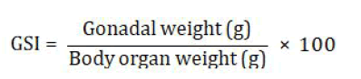
Gonadosomatic Index (GSI)
GSI was calculated at the end of the experiment for each animal by using the formula given below.
Relative organ weight
Relative weight of organ of each rat was calculated by using the formula given below.

Biochemical analysis
Testicular tissues of treated and control rats were examined for antioxidant enzymes analysis. Homogenate of testicular tissue was prepared with the help of automatic homogenizer machine by taking 90 mg tissue in 1 mL phosphate buffer saline with pH of 7.4. Homogenate was centrifuge with 13000 rpm for 30 mins. After centrifugation, the supernatant was isolated for the measurement of oxidative stress.
Catalase (CAT)
For the measurement of CAT level, 0.1 mL of homogenate was diluted in 2 ml of phosphate buffer (pH 5.0) along with 0.4 ml H2O2 (5.9 nM) in the cuvette. After thoroughly mixing, the absorbance was read with spectrophotometer having 240 nm wavelength with a time interval of 15 s and 30 s. One unit of CAT was defined as a change in the absorbance of 0.01 units in one mint.
Peroxidase (POD)
The activity of POD is estimated by using protocol of Calberg and mannervik, 1975 with little modifications. Homogenate was taken 0.1 mL and mixed with 0.1 mL of guaicol which was 20 mM. After that 0.3 mL of H2O2 and 2.5 mL phosphate buffer (pH=5.0) was mixed in the solution. Absorbance change was measured with the help of spectrophotometer at 470 nm. One unit of enzyme activity was changed in absorbance of 0.01 per minute.
Superoxide Dismutase (SOD)
The SOD activity was measured by the method. Homogenate was taken 0.3 mL and it was mixed with 0.1 mL of phenazine methosulphate and 1.2 mL of sodium pyrophosphate with pH 7.0 was added in it. The reaction started after addition of the 0.2 mL NADH and stopped after addition of 1 mL glacial acetic acid. Chromogen formed was measured with the help of spectrophotometer at 560 nm absorbance.
Thiobarbituric Acid Reactive Substances (TBARS)
TBARS assay is usually used for the detection of Malondialdehyde (MDA) which is the major product of lipid peroxidation. This assay was performed according to the procedure given by Ohkawa and Iqbal 1996. Homogenate, 0.1mL was added to the 0.29 mL of 0.1 M phosphate buffer and 0.1 mL of ascorbic acid and incubated on 37°C in a shaking water bath for 1 hour. Reaction stopped after addition of 0.5 mL of 10% trichloroacetic acid. After that 1 mL of trichlorobarbituric acid (0.67%) was added and test tubes were put in hot water for 20 minutes at 95°C. After boiling test tubes were shifted to crushed ice box for 20 minutes. After chilling for 20 minutes in the crushed ice, material was centrifuged at 30,000 rpm for 10 minutes. The absorbance of the supernatant was measured with the help of spectrophotometer at 535 nm and result was nm TBARS/mint/mL.
Reactive Oxygen Species (ROS)
Amount of ROS in the sample was measured with the help of protocol given by Hayashi et al. Homogenate, 5 μL was added to the 140 μL sodium acetate buffer (pH=4.8) in the microtiter 96 wells plate and incubated for 5 minutes on 37â??. DEPPD and ferrous chloride solution were taken in a 1:25 ratio (R1, R2). This solution of R1+R2 was put in each well and incubated for 1 minute at room temperature. Absorbance was measured on 505nm after 15 seconds of interval for 2 minutes.
A standard curve was plotted and the ROS value was measured from it.
Analysis of plasma hormonal level
Plasma was separated from the blood by centrifugation on 3000 rpm for 15 minutes and used for hormone analysis of FSH, LH, estradiol, progesterone and testosterone with the help of Enzyme-Linked Immunosorbent Assay (ELISA) kit. The assay was done according to the instruction provided with the kit.
Lipid profile
Plasma level of High-Density Lipoprotein (HDL) and Low- Density Lipoprotein (LDL) was determined with the help of specific enzymatic kit (AMP diagnostic kit) on chemical analyzer by following the instruction provided by the manufacturer on the kit.
Tissue histology
Testes were fixed in formalin (10%) for forty-six to forty-eight hours. After fixation, testes were removed out from formalin and passed from different grades of ethanol for dehydration. Then tissue was processed in xylene until they become clear. Tissue was then put in paraffin wax and 7 μm thick sections were cut with the help of microtome. Sections were stained in eosin and hematoxylin by following the standard protocol. After staining, slides of all groups were observed under light microscope on 40X. Photographs of the tissue sections were captured with the help of Leica LB microscope (Germany) equipped with a camera (Canon Japan). For histomorphometric analysis, images were captured at 40X and twenty images per animals were selected for the calculations of seminiferous tubule diameter, lumen diameter and epithelial height by using the software image J2x.
Statistical analysis
For the analysis of data, one-way ANOVA test was run followed by Dunnet multiple comparison test for the comparison of test groups with control on Graph Pad Prism version 5 with probability value less than 0.05 (P<0.05).
Ethics approval and consent to participate
The study was conducted in Koc University Graduate School of Health Sciences. All methods were performed in accordance with relevant guidelines and regulations of “Local Ethics Committee for Animal Experiments of Koc University.” The animals were kept in the Koc University, Animal Research Facility (KUARF) of Centre for Translational Medicine (KUTTAM). The study was approved by the committee with Approval No. (2022- 15).
Results
Body weight
Effect of daily consumption of broiler and domestic chicken meat on body weight and weight gain in the male rat. Mean body weight of each group was calculated at PND 0, PND 21, PND 60 and PND 90 (Table 1). There was no significant difference between control and meat fed groups at PND 0 and PND 21. At PND 60, there was a significant increase in weight of B1 group (P<0.05), B2 group (P<0.01) and D2 group (P<0.05). At PND 90, B1 and B2 (P<0.01) and D2 (P<0.05) showed a signi icant increase in weight as compared to control. Weight gain was high in B1 (P<0.05), B2 (P<0.01) and D2(P<0.05). There was no signi icant difference between D1 and control.
| Body weight (g) |
| Groups n=5 |
PND 0 |
PND 21 |
PND 60 |
PND 90 |
Weight gain |
| Control |
5.45 ± 0.26 |
37.80 ± 1.47 |
197.40 ± 2.44 |
241.80 ± 3.18 |
204.00 ± 3.00 |
| B1 |
5.47 ± 0.21 |
39.20 ± 1.72 |
210.20 ± 2.78* |
258.40 ± 3.44** |
219.20 ± 2.52* |
| B2 |
5.60 ± 0.23 |
36.00 ± 1.59 |
212.20 ± 3.40** |
259.40 ± 2.79** |
223.40 ± 4.15** |
| D1 |
5.52 ± 0.15 |
38.20 ± 1.72 |
203.60 ± 2.69 |
250.20 ± 2.52 |
212.00 ± 2.47 |
| D2 |
5.49 ± 0.18 |
36.60 ± 1.51 |
208.60 ± 2.44* |
253.60 ± 2.91* |
217.00 ± 4.06* |
Table 1: Comparative alteration in body weight of male rats after chronic consumption of broiler and domestic chicken meat for 90 days. Results of body weight and weight gain are expressed in Mean ± SEM.
Effect of daily consumption of broiler and domestic chicken meat on GSI paired testicular weight, absolute and relative epididymal weight, testes longitudinal and transverse diameter
Mean GSI, absolute and relative weight of the testes and epididymis was calculated on PND 90 after chronic consumption of chicken meat (Table 2). GSI showed non-significant difference between control and experimental group. There was nonsignificant difference in weight of the testes, longitudinal and transverse diameter between control and experimental group. The absolute epididymal and relative epididymal weight of 0.34 g broiler chicken meat fed was signi icant increased compared to 0.34 g domestic chicken meat consuming rats.
| Parameters |
Groups |
| Control |
B1 |
B2 |
D1 |
D2 |
| Gonadosomatic index |
0.97 ± 0.02 |
0.93 ± 0.04 |
0.91 ± 0.05 |
0.95 ± 0.01 |
0.92 ± 0.02 |
| Paired testes weight (g) |
2.35 ± 0.03 |
2.39 ± 0.10 |
2.35 ± 0.12 |
2.37 ± 0.03 |
2.33 ± 0.04 |
| Absolute epididymal weight (g) |
1.18 ± 0.04 |
1.07 ± 0.03 |
1.92 ± 0.03* |
1.13 ± 0.08 |
1.06 ± 0.06 |
| Relative epididymal weight |
4.88 ± 0.23 |
4.14 ± 0.12 |
5.90 ± 0.15** |
4.50 ± 0.31 |
3.0 ± 0.19* |
| Longitudinal diameter of tests |
10.40 ± 0.49 |
9.83 ± 0.40 |
10.12 ± 0.30 |
11.21 ± 0.49 |
10.91 ± 0.80 |
| Transverse diameter of tests |
6.66 ± 0.46 |
6.30 ± 0.46 |
7.09 ± 0.43 |
7.40 ± 0.40 |
6.63 ± 0.45 |
| Seminal vesicle weight (g) |
0.93 ± 0.03 |
0.91 ± 0.02 |
1.5 ± 0.03** |
0.64 ± 0.02 |
0.70 ± 0.03* |
| Prostate weight (g) |
0.66 ± 0.02 |
0.67 ± 0.03 |
0.94 ± 0.05** |
0.37 ± 0.05 |
0.72 ± 0.02* |
| Note: Results of GSI and organ weight are expressed in Mean ± SEM. *P<0.05, **P<0.01. |
Table 2: GSI, reproductive organ weight and testicular diameter of rats after chronic consumption of broiler and domestic chicken meat for 90 days.
Accessory reproductive organ weight
Effect of chronic consumption of broiler and domestic chicken meat on the weight of seminal vesicle and prostate
Mean weight of seminal vesicle and the prostate gland was calculated on PND 90 after chronic consumption of chicken meat (Table 2). Weight of seminal vesicle and prostate gland in the B2 meat fed groups was increased significantly compared to control and D2.
Weight of body organs
Effect of chronic consumption of broiler and domestic chicken meat on the weight of Kidney, heart and liver.
Biochemical analysis
Mean weight of organs kidney, heart and liver were calculated on PND 90 (Table 3). experimental groups.
There was significant variation between weight of kidney and liver in control and the control. There was no effect of B1 and D1 meat fed group on kidney and liver weight. Kidney and liver weight were significantly increased in B1 (P<0.05) and B2 (P<0.01) groups compared to weight compared to control D1 and D2 group. D1 group, liver weight did not vary significantly compared to control. Similarly, weight of heart was compared with control and experimental group. Non-significant difference was found among these groups.
| Groups (n=5) |
Kidney |
Heart |
Liver |
| Control |
1.02 ± 0.04 |
1.02 ± 0.05 |
7.76 ± 0.46 |
| B1 |
1.23 ± 0.08 |
1.04 ± 0.04 |
8.92 ± 0.24* |
| B2 |
1.39 ± 0.02** |
1.02 ± 0.03 |
9.92 ± 0.25** |
| D1 |
1.24 ± 0.09 |
1.06 ± 0.02 |
8.0 ± 0.23 |
| D2 |
1.25 ± 0.05* |
1.04 ± 0.03 |
8.5 ± 0.14 |
Table 3: Kidney, heart and liver weight of male rats after chronic consumption of broiler and domestic chicken meat for 90 days. Results of body organ weight are expressed in Mean ± SEM. *P<0.05.
Oxidative stress
Effect of chronic consumption of broiler and domestic chicken meat on antioxidant enzyme level in the testes. The mean level of oxidative stress markers in testes was measured on PND 90 (Table 4).
| |
Control |
B1 |
B2 |
D1 |
D2 |
| CAT (U/mg protein) |
7.38 ± 0.47 |
6.20 ± 0.95 |
5.96 ± 0.86 |
6.98 ± 0.36 |
7.12 ± 0.37 |
| SOD (U/mg protein) |
25.52 ± 2.50 |
21.86 ± 1.49 |
22.25 ± 2.00 |
24.47 ± 1.55 |
22.07 ± 1.56 |
| POD (nmole) |
14.14 ± 1.38 |
12.20 ± 1.57 |
10.89 ± 1.89 |
13.87 ± 0.79 |
11.05 ± 2.02 |
| TBARS (nM/TBAR/min/mgprotein) |
16.76 ± 1.63 |
19.16 ± 0.95 |
17.90 ± 0.89 |
16.79 ± 0.77 |
17.30 ± 0.95 |
| ROS (U/mg tissue) |
0.82 ± 0.02 |
0.85 ± 0.03 |
0.94 ± 0.04* |
0.81 ± 0.02 |
0.86 ± 0.02 |
| HDL (mg/dL) |
60.13 ± 2.57 |
56.44 ± 2.63 |
53.08 ± 2.82 |
59.09 ± 2.50 |
58.08 ± 2.66 |
| LDL (mg/dL) |
40.94 ± 2.82 |
44.88 ± 2.38 |
48.76 ± 2.94* |
41.62 ± 3.72 |
41.99 ± 3.79 |
| Testosterone (nM/L) |
6.54 ± 0.49 |
5.04 ± 0.39 |
7.64 ± 0.43* |
6.79 ± 0.40 |
5.42 ± 0.49 |
| Estradiol (pg/mL) |
7.30 ± 0.49 |
9.60 ± 0.74 |
10.38 ± 0.26* |
7.44 ± 0.40 |
7.93 ± 1.22 |
| Progesterone (ng/mL) |
4.31 ± 0.70 |
3.97 ± 0.49 |
3.49 ± 0.58 |
4.30 ± 0.50 |
4.12 ± 0.61 |
| LH (mU/L) |
3.39 ± 0.8 |
3.41 ± 0.5 |
5.95 ± 0.7* |
3.31 ± 0.4 |
3.85 ± 0.9 |
| FSH (mU/L) |
34 ± 9.5 |
36 ± 0.2 |
38 ± 0.4* |
34 ± 0.1 |
36 ± 9.0 |
Table 4: Comparative antioxidant enzyme, plasma HDL, LDL and plasma steroidal hormone level of male rats a ter chronic consumption of broiler and domestic chicken meat for 90 days. Results are expressed in Mean ± SEM. *P<0.05.
Assays were done to detect the level of antioxidant enzymes, ROS and lipid peroxidation in testes. Level of antioxidant enzymes did not vary significantly between control and test groups. Similarly, effect on lipid peroxidation was detected significantly in any group. However Reactive oxygen species level increased significantly (P<0.05) in the testes of B2 group. ROS level was not significant also elevated in B1 and D2 groups.
Hormonal analysis
Effect of chronic consumption of broiler and domestic chicken meat on plasma level of testosterone, estradiol and progesterone.
The concentration of testosterone, estradiol and progesterone were measured in the blood plasma on PND 90 (Table 4).
The plasma testosterone and ROS level was significantly increased in B2 compared to control, D1 and D2 droups. Nonsignificant difference was observed in the progesterone level. However, in the B2 group, estradiol level increased significantly (P<0.05) as compared to the control group.
Lipid profile
Effect of chronic consumption of broiler and domestic chicken meat on High-Density Lipoprotein (HDL) and Low-Density Lipoprotein (LDL).
Level of HDL and LDL in blood was measured on PND 90 following chronic consumption of broiler and domestic chicken meat (Table 4).
Level of HDL was low in B2 group, but this decrease in HDL level was not found significant compared to control. Similarly, LDL level was comparatively high in B2 groups compared to control and D2.
Histomorphometric analysis
Effect of chronic consumption of broiler and domestic chicken meat on seminiferous tubule diameter, epithelial height and lumen diameter.
Seminiferous tubular diameter, the height of epithelial layer and lumen diameter of seminiferous tubule was measured with Image J2 software and data was presented in Figure 1.
Morphologically multiple vacuoles and altered grass morphology of seminiferous tubule was observed in B2 and were less prominent in other experimental groups. However seminiferous tubule diameter, the epithelial height of the tubule and lumen diameter of experimental groups were compared with control. In rats B2 fed with chronic broiler meat showed a marked decreased seminiferous tubule diameter, decreased epithelial height and increased lumen diameter changes that were more significant compared to rats fed with D2 domestic chicken meat.

Figure 1: Photomicrograph (40X) of testes showing seminiferous tubule, seminiferous tubular diameter, lumen diameter and height of epithelial cells. (A) Control, (B) Seminiferous tubule of rat fed with B1 broiler meat, (C) Seminiferous tubule of rat fed with B2 broiler meat showing reduced epithelial height increased lumen diameter and low sperm quantity in lumen as compared to control, (D) Seminiferous tubule of rat fed with D1 domestic chicken meat, (E) Seminiferous tubule of rat fed with D2 domestic chicken meat.
Discussion
Dietary patterns influence risk for obesity and consequently endocrine disorders. With increasing concerns related to the use of red meat which contains saturated fatty acid, broiler chicken has become a priority among the mass population. Tremendous genetic selection for fast growth rate and feed conversion was carried out to maximize the size of broiler [19]. Various fats in the form of lard, tallow, yellow grease, saturated fats from animal sources are added in the broiler feed to fulfill their energy requirement [20]. There is speculation that their residues become impregnated in the form of body fat. Broiler which was considered as low-fat food is no longer a lean one. More energy from consumption of broiler is through fat than from protein with the reduction of omega-3 fatty acid and this has likely to be an adverse effect on health. As we know, the consumption of high-fat energy diet is a risk factor for the development of obesity and type 2 diabetes. Moreover, high-fat energy diet causes alteration in the Kiss1 and GPR54 which account for metabolic and reproductive abnormalities in obese individuals.
In our study we investigated the comparative effect of chronic consumption of domestic chicken meat and broiler commercially available meat on reproductive system, testicular morphology, sexual hormones and oxidative stress profile in postnatal male rats. Rats were chronically fed on two different quantities of low and high quantity of chicken and domestic meat B1, D1 (0.17 g) and B2, D2 (0.34 g). There was significant increase in body weight and weight gain in B2 compared to D40 group. In gonadosomatic index absolute and relative epidydimal weight, weight of seminal vesical and prostate weight was significantly increased in B2 compared to control and D2. Evident increase in kidney and liver weight in B1, B2 and nonâ?significant increase in D2 groups. Level of reactive oxygen species in B2 was significantly higher than control D2 and other groups. Serum level of FSH, LH, testosterone, estradiol and low density lipoprotein in B2 was significantly increased compared to control and D2. In B2 rats fed with 0.34 g broiler meat showed a marked decreased seminiferous tubule diameter, decreased epithelial height and increased lumen diameter changes that were more significant compared to rats fed with 0.34 g domestic chicken meat. Meat was found to be exerting a significant effect on weight gain with most pronounced effect with the broiler meat consumption. This increase in body weight could be due to consumption of fats and cholesterol in the meat which provides precursor for the formation and storage of fat in the adipose tissues. Broiler chicken is reared on feed which contains large ratio of cholesterol and fats which enables broilers to gain weight and to fulfill its fast-growing energy requirement and these fats get deposited in the meat without much modification. Similar results were found in the study of Ahmad et al., which documented that commercial broiler chicken consumption had significant effect on the weight gain in the female rat. A research carried by Komprda et al. stated that commercially reared chicken without the skin represents the 55% of upper limit of daily cholesterol intake. In our study rats fed with B2 showed increased body weight and significant elevated level of LDL and less HDL. Weight increase was observed after postnatal day 60. Similar results were presented by Ullah et al., which showed that rats that are fed with high-fat diet had increased body weight and this effect was more intense in male than female but highfat diet did not affect the weight of the pups through pregnant and lactating mothers showed that the type of meat consumption affects body composition in adolescence. He found that high poultry meat intake at 10 years of age increases the fat mass index of the body. Our results are contrary to the Van Hecke et al., which showed that feeding the rats with white chicken and red beef meat did not have a significant effect on weight gain throughout the experimental feeding period. This difference in weight gain can be due to different rearing conditions of model animal, different strains of chicken species and difference in fat content in meat due to different feed used.
In our research, there was no significant effect on the weight of testes and marked variation was found in absolute and relative weight of epididymis similar to prostate and seminal vesicles. Inconsistent with our observations, Ahmad et al., found that broiler meat had a significant effect on the reproductive organ of the female rat. After consumption of meat, ovary weight increased significantly and appearance of the ovary was swollen and relatively cystic as compared to control [1]. The previous study had shown that diet rich in lard/saturated fatty acid effect the testes morphology and increased the fat mass of epididymis but this result was not replicated in our study.
With increasing prevalence of obesity, fatty liver disease is growing, and diet plays very important role in its development. Consumption of high-fat diet leads to an increase in the weight of the liver because of the great delivery of FAs to the liver which exceeds the removal capacity. Our result showed the increase in the weight of liver after consumption of both types of meats in the diet. This can be due to high-fat content in the meat of broiler and overâ?consumption of protein in the domestic chicken which considered as the lean meat an important source of animal protein. This increase in liver weight could be due to high acid load and deposition of triglycerides leading to inflammation of the liver. Our results were in contradiction to which documented that diet with high animal protein reduced the liver fat and weight. However, provided the data that high protein increased the liver inflammation. Our results documented the increase in weight of the kidney in case of high quantity of broiler chicken meat compared to domestic chicken. This increase in kidney weight could be due to an increase in the waste product after protein metabolism.
Lipid present in the diet gives rise to the formation of the lipids oxidation product such as malondialdehyde. Effect on the presence of MDA and oxidative stress in the reproductive organ due to meat consumption has not been studied so far. This may be the first time that oxidative stress on reproductive organs due to the consumption of white meat was analyzed. In our result, it was found that antioxidant enzymes nonâ?significantly decreased in the testes and MDA level increased nonsignificantly, but the only significant effect was the increase in the level of ROS in the testes of broiler fed group. This increase in ROS level could be due to high body weight as there is a positive relationship between body mass and ROS level in the testes. High fat and cholesterol content in broiler meat may be accumulated in the body and increased the ROS level in testes. If spermatogenesis is impaired due to obesity, proper cytoplasm does not extrude and level of ROS increases due to activation of NADPH oxidase. This increase in ROS can be of exogenous nature, due to fat-soluble toxins in the meat of broiler. Some previous studies have provided the effect of meat consumption of oxidative stress level in stomach, liver, and plasma. Provided the data that red meat and lard mixed chicken meat increases the lipid oxidation product; malondialdehyde, hexanal and 4 hydroxy-2-neonatal in the blood and stomach of the rat and decreased the glutathione level. However, in another study it was observed that red meat did not have any effect on the oxidative stress level or inflammation of the body.
In the previous study documented showed that rats had significantly high cholesterol level when they were fed on broiler chicken meat. In our result, we found that in broiler fed group there was an increase in the level of LDL and decrease in the level of HDL although the effect was not significantly different. Previously it was stated that red meat high in saturated fatty acid had adverse effect on the body cholesterol level increases the risk factor for cardiovascular diseases. White meat especially chicken was considered as lean meat source enriched with polyunsaturated fatty acid. However, the fat that is high in saturated fatty acids is added to the broiler feed which deposited in the muscle which modification because broiler in monogastric animals and is affecting the composition of broiler fat. So fat and cholesterol residue in the broiler meat can provide a precursor for the increase cholesterol level of the body [14]. The increase in cholesterol level may result in an increase in the synthesis of steroid hormones as cholesterol is the precursor for the steroid hormone leading to imbalance of hormones in the body affecting the HPG axis.
In our study, it was observed that consumption of broiler meat leads to steroid hormonal imbalance in the rat plasma with significantly increased testosterone and estradiol level and no significant effect on the progesterone level. There was no significant difference in the hormone level in other dietary groups. This hormonal imbalance is enlightened that hypothalamic pituitary testicular axis was disturbed. Same result was seen which showed that broiler meat increased the estradiol level and decrease the progesterone level in female rat. In her study, level of testosterone increased results are inline to our as level of testosterone significantly increased.
They found that a high fat diet increases the estrogen level in body and decreases the progesterone level in the female rat. Variation in the steroidal sex hormones could be due to concentrates of fat and cholesterol in the flesh of broiler from the feed they used and consumption of this meat resulted in augmented production of steroid hormones from cholesterol in the body. They showed that increase LDL or HDL increased the production of steroidal sex hormone in differentiating adrenocortical cells. In male testosterone is converted into estrogen with the help of aromatase enzyme and activity of aromatase enzyme increases by increasing the adipose tissue, so production rate of estrogen relates closely to the body weight. High circulating estradiol may cause the inhibition of the HPG axis resulting in low GnRH and LH secretion which ultimately decreases the testosterone production from gonads. In broiler fed group body weight was increased and leptin level increases with an increase in body fats. Increase in leptin level had a negative correlation with the level of testosterone. Leptin either indirectly modulates the production of testosterone through kiss 1 gene expression of directly by blocking the function of Leydig cells by inhibiting the conversion of progesterone into testosterone.
In rats fed chronically with broiler meat showed a marked decreased seminiferous tubule diameter, decreased epithelial height and increased lumen diameter changes that were more significant compared to rats fed with high dose of domestic chicken meat. Weight of seminal vesicle and prostate gland in the B2 meat fed groups was increased significantly compared to control and D2.
Conclusion
Traditionally, chicken meat was one of the important sources of protein and essential fatty acid but nowadays, broiler husbandry practices along with the use of saturated fats from different rendering sources affect the meat quality by increasing the fat energy content. This high fat energy content meat adversely affects the male reproductive system by either providing a source of exogenous steroids hormone or cholesterol which could interfere with the steroidogenesis pathway of the male reproductive system or through the development of obesity which transduce the metabolic information to the hypothalamic pituitary testicular axis leading to hormonal imbalance and increased oxidative stress.
Author Contribution
I hereby declare that the work presented in the article is my own effort, and that the article is my own composition. No part of this article has been previously published in any journal.
Funding
Funding source was not available.
Availability of Data and Materials
All data generated or analyzed during this study are included in this article.
Arrive Guidelines
The study is reported in accordance with arrive guidelines.
Conflict of Interest
The authors declare that the research was conducted in the absence of any commercial or financial relationships and declared no conflict of interest.
Consent for Publication
Not applicable.
Ethical Approval
The study was conducted in Koç University graduate school of health sciences. All methods were performed in accordance with relevant guidelines and regulations of “Local Ethics Committee for Animal Experiments of Koc University.” The animals were kept in the Koç University, Animal Research Facility (KUARF) of Centre for Translational Medicine (KUTTAM). The study was approved by the committee with Approval No. (2022-15).
References
- Ahmad S, Ahmed I, Haider S, Batool Z, Ahmed F, et al. (2018) Effects of consumption of caged and un-caged chicken meat on ovarian health of female Wistar rats. Pak J Zool 50.
[Google Scholar]
- Al-Nasser A, Al-Khalaifa H, Al-Saffar A, Khalil F, Albahouh M, et al. (2007) Overview of chicken taxonomy and domestication. J World's Poult Sci 63: 285-300.
[Crossref] [Google Scholar]
- Memon IN, Noonari S, Asif M, Shah ST, Peerzado MB, et al. (2015) Economic analysis of poultry egg production in Quetta District Balochistan. J Fish Lives 3: 2332-2608.
[Crossref] [Google Scholar]
- Knowles TG, Kestin SC, Haslam SM, Brown SN, Green LE, et al. (2008) Leg disorders in broiler chickens: prevalence, risk factors and prevention. PloS One 3: e1545.
[Crossref] [Google Scholar]
- Julian RJ (1998) Rapid growth problems: Ascites and skeletal deformities in broilers. Poult Sci 77: 1773-1780.
[Crossref] [Google Scholar] [PubMed]
- Ortiz MR, Valdivia FA, Martínez RJ, Martínez de AA (2000) Effect of clenbuterol on growth performance in broilers. 52: 256-260.
[Google Scholar]
- Buttery PJ, Dawson JM (1987) The mode of action of beta-agonists as manipulators of carcass composition.
[Google Scholar]
- Cardoso LA, Stock MJ (1996) Effect of clenbuterol on growth and body composition during food restriction in rats. J Anim Sci 74: 2245-2252.
[Crossref] [Google Scholar] [PubMed]
- Schiavone A, Tarantola M, Perona G, Pagliasso S, Badino P, et al. (2004) Effect of dietary clenbuterol and cimaterol on muscle composition, βâ?adrenergic and androgen receptor concentrations in broiler chickens. J Anim Physiol Anim Nutr 88: 94-100.
[Crossref] [Google Scholar] [PubMed]
- Yousefi J, Maheri-Sis N, Shaddel-Telli A, Hatefinezhad K, Eshartkhah B, et al. (2011) Effect of salbutamol (a beta-adrenergic agonist) on growth performance of broiler chickens.
[Google Scholar]
- Lu H, Zhang H, Zhu T, Xiao Y, Xie S, et al. (2017) Metabolic effects of clenbuterol and salbutamol on pork meat studied using internal extractive electrospray ionization mass spectrometry. Sci Rep 7: 5136.
[Crossref] [Google Scholar] [PubMed]
- Malucelli A, Ellendorff F, Meyer HHD (1994) Tissue distribution and residues of clenbuterol, salbutamol, and terbutaline in tissues of treated broiler chickens. J Anim Sci 72: 1555-1560.
[Crossref] [Google Scholar] [PubMed]
- Rondelli SG, Martinez O, Garcia PT (2004) Effects of different dietary lipids on the fatty acid composition of broiler abdominal fat. Braz J Poult 6: 171-175.
[Crossref] [Google Scholar]
- Ahmad S, Ahmed I, Haider S, Batool Z, Ahmed SB (2017) Daily consumption of commercial chicken feed and meat lead to alterations in serum cholesterol and steroidal sex hormones in female rats. Pak J Pharm Sci 30.
[Google Scholar] [PubMed]
- Ahmad S, Omm-e-Hany AI, Ahmed SA, Alamgir A, Neelam A (2016) Potential effect of chicken boneless meat on the body weight and serum cholesterol levels of the female Albino Wister rats: In direct human prospective studies. Am Eurasian J Agric Enviorn Sci 16: 466-469.
[Google Scholar]
- Saara Ahmad SA, Iftikhar Ahmed IA, Saida Haider SH, Zehra Batool ZB, Fatima Ahmed FA, et al. (2018) Effects of consumption of caged and un-caged chicken meat on ovarian health of female wistar rats.
[Google Scholar]
- Ahmad S, Ahmed I (2014) Response of Wistar rats to broiler chicken feed and soybean on body weight, obesity and weight of selected visceral organs. Pak J Biochem Mol Biol 47: 137-140.
[Google Scholar]
- Emmerson DA (1997) Commercial approaches to genetic selection for growth and feed conversion in domestic poultry. Poult Sci 76: 1121-1125.
[Crossref] [Google Scholar] [PubMed]
- Firman JD, Kamyab A, Leigh H (2008) Comparison of fat sources in rations of broilers from hatch to market. Int J Poult Sci 7: 1152-1155.
[Google Scholar]
- Wang Y, Lehane C, Ghebremeskel K, Crawford MA (2010) Modern organic and broiler chickens sold for human consumption provide more energy from fat than protein. Public Health Nutr 13: 400-408.
[Crossref] [Google Scholar] [PubMed]
Citation: Aslam M (2025) A Comparative Analysis of Hypothalamic Pituitary Gonadal Axis Alterations by Chronic Consumptions of Broiler and
Domestic Chicken Meats in Postnatal Sprague Dawley Male Rats. Health Sci J Vol.19 No.2

