Flyer

Health Science Journal

  • ISSN: 1108-7366
  • Journal h-index: 51
  • Journal CiteScore: 10.69
  • Journal Impact Factor: 9.13
  • Average acceptance to publication time (5-7 days)
  • Average article processing time (30-45 days) Less than 5 volumes 30 days
    8 - 9 volumes 40 days
    10 and more volumes 45 days
Awards Nomination 20+ Million Readerbase
Indexed In
  • Genamics JournalSeek
  • China National Knowledge Infrastructure (CNKI)
  • CiteFactor
  • CINAHL Complete
  • Scimago
  • Electronic Journals Library
  • Directory of Research Journal Indexing (DRJI)
  • EMCare
  • OCLC- WorldCat
  • University Grants Commission
  • Geneva Foundation for Medical Education and Research
  • Euro Pub
  • Google Scholar
  • SHERPA ROMEO
  • Secret Search Engine Labs
Share This Page

Research Article - (2023) Volume 17, Issue 10

Efficacy of Taxol and Prunella Vulgaris against human lung cancer cell line-A549.

Darakhshanda Neelam1*, S.A. Husain2, Tanveera Tabasum3 and M. Maqbool lone4
 
1Department of Advance Centre for Human Genetics, SKIMS, Srinagar, Kashmir, India
2Jamia Millia Islamia, New Delhi, India
3Department of Zoology, Cluster University, M.A. road, Srinagar, Kashmir, India
4Department of Radiation Oncology, SKIMS, Srinagar, Kashmir, India
 
*Correspondence: Darakhshanda Neelam, Department of Advance Centre for Human Genetics, SKIMS, Srinagar, Kashmir, India, Tel: 9906455633, Email:

Received: 10-Oct-2023, Manuscript No. Iphsj-23-14093; Editor assigned: 12-Oct-2023, Pre QC No. Iphsj-23-14093(PQ); Reviewed: 25-Oct-2023, QC No. Iphsj-23-14093; Revised: 28-Oct-2023, Manuscript No. Iphsj-23-14093(R); Published: 31-Oct-2023

Abstract

Introduction: Medicinal plants play a central role in the healthcare system of the large proportions of the world’s population. Plant derived compounds are an important source of several clinically useful anticancer agents. Cytotoxicity screening models are the preliminary methods for the selection of active extracts against cancer.

Materials and methods: A comprehensive study was carried out to determine the Cytotoxic activity of active extracts of the plant-P. vulgaris, T. buccatta against human lung cancer (A549) cell line at eight different concentration to determine the IC50 (50% growth inhibition) by MTT assay including morphological study by fluorescence microscopy, Apoptosis by Annexin V, caspase 3-7 assay and levels of expression of genes. Each sample was assayed in triplicate and control samples include cells without plant extracts.

Results: Results demonstrate that the percentage of growth inhibition increases with increasing concentrations of test compounds. The P. vulgaris extracts induced significant cytotoxic effects on the A549 cancer cell line and these effects were stronger than the other selected plant extracts reflecting a promising way of chemotherapy to counter the spread of non- small cell lung cancer.

Conclusion: Extract which exhibit substantial antipoliferative and apoptic activity may represent a source for novel natural anticancer entities.

Keywords

A549; P. vulgaris; T. buccatta

Introduction

Over the decades, herbal medicine has become a topic of global importance, making an impact on both world health and international trade. Medicinal plants continue to play a central role in the healthcare system of large proportions of the world’s population. This is particularly true in developing countries, where herbal medicine has a long and uninterrupted history of use. Recognition and development of the medicinal and economic benefits of these plants are on the increase in both developing and industrialized nations. Continuous usage of herbal medicine by a large proportion of the population in the developing countries is largely due to the high cost of western pharmaceuticals and healthcare.

Among the human diseases treated with the medicinal plants is cancer. Globally, cancer represents a substantial burden of disease in the community and appears to be a prime cause of concern. Cancer is a third leading cause of mortality and it strikes more than the one third of world’s population and it is the cause of more than 20% of all deaths [1]. Cancer is depicted as a disease when cell proliferation and differentiation becomes uncoupled and damage to numerous regulatory genes resulting in the development of invasive and metastatic cancer which is the culmination of the chronic disease process, carcinogenesis [2]. According to the International Agency for Research on Cancer (2002), cancer killed >6.7 million people around the world and another 10.9 million new cases were diagnosed [3]. If the results are extrapolated, at the same rate, an estimated 15 million people will have cancer, annually, by 2020. According to an estimate given by American Cancer Society (2009), about 1,500,000 new cases and over 500,000 deaths are expected in the US by 2009. Every year over 200,000 people are diagnosed with cancer in the United Kingdom only, and approximately 120,000 die as an aftermath of the disease (Department of Health, 2000). The National Cancer Registry of South Africa has spotted the cancers of bladder, colon, breast, cervix, lungs and melanoma commonly among inhabitants [4]. In India around 55000 people died of cancer in 2010, according to eternals in the Lancet today (March 2012) as stated by the International Agency for Research on cancer (IARC). Parts of India have the world’s highest incidence of cancers of gall bladder, mouth and lower pharynx, India’s first cancer atlas shows. Among the states of India, J&K also shows the highest incidence of cancer [5-8 ].

Among this dreadful disease Lung cancer is the leading cause of cancer deaths and its incidence continues to rise worldwide. Small-cell lung cancer makes up to about 25% of lung cancer cases and is characterised by a rapid and aggressive clinical course. Any practical solution in combating this dreadful disease is of paramount importance.

Taxanes are the most recently solicited chemotherapeutic drugs of our era. During the past decades, these unique hydrophobic mitotic inhibitors have been thoroughly investigated through numerous experimental and clinical trials which have brought hope in breast, ovarian, lung, prostate [9 ,10], pancreas, gastric [11,12] and head and neck [13] cancer treatments.

In brief, the taxanes mainly group Paclitaxel (Taxol) and Docetaxel (Taxotere) as well as taxanes homologs, which are derived from natural sources; taxol [14] is derived originally from Tagus Brevifolia (bark of Pacific yew/Western yew conifers) while Docetaxel is a semisynthetic analogue of the latter; an esterified derivative of 10-deacetylbaccatin-III (10-DAB) extracted from Taxus Baccata [15 ] (needles of European yew tree). During course of time due to poor oral bioavailability, solubility and numerous side effects; views for the development of new similar antimitotic have been encouraged and brought to light. Moreover, since there have been numerous multidrug resistance (MDR) [16] in patients, combination therapies are preferred over single drug therapy and Texans also known to be having a radio sensitizing [13] effect have proved to be helpful in the palliative treatment of patients if not in the curative one. After an initial response to chemotherapy, most small cell lung cancers relapse in a drug resistance form, leading to a 2 year survival of only 5% [1]. The emergence of resistance to chemotherapeutic agents remains a major problem in the treatment of patients with small-cell lung cancer, despite the fact that these patients usually have a good initial response to chemotherapy. Although a number of genetic alterations associated with drug resistance in small-cell lung cancer are known, including the deregulated expression of proteins involved in drug transport and activity [17], and in cell cycle checkpoint control [18], few advances have been made in improving the therapeutic options.

Despite, their side effects related mostly to their vehicles [19], they remain one of the most acceptable treatments for metastatic breast [20], ovarian, prostate and lung carcinomas. The list for laboratory experiments and clinical oncological trials is very long concerning taxanes; but their outcome is of tremendous eagerness in the cancer field. An alternative solution to western medicine embodied with severe side effects is the use of medicinal plant preparation to arrest the insidious nature of the disease. Attempts are underway to work out the therapeutic and anti-neoplastic properties of medicinal plants [21-26]. A recent review of Butler lists seventy natural products or natural product derivatives currently undergoing trials in United States, Europe, Japan and Korea [27 ]. Out of seventy compounds twenty eight were plant derived.

There are about 45,000 plant species in India, with concentrated hotspots in the region of Eastern Himalayas, Western Ghats and Andaman & Nicobar Island. The officially documented plants with medicinal potential are 3000 but traditional practitioners use more than 6000. India is the largest producer of medicinal herbs and is appropriately called the botanical garden of the world [28]. There are currently about 250 000 registered medical practitioners of the Ayurvedic system (total for all traditional systems: approximately 291 000), as compared to about 700,000 of the modern medicine system. In rural India, 70 per cent of the population is dependent on the traditional system of medicine, the Ayurveda [29]. In India, the J&K region is endowed with a wide range of physiography, climate, soil and biological wealth. The region is one of the richest reservoirs of biological diversity in the world and is considered as a store house of the valuable medicinal plant species [30,31]. Cytotoxicity screening models are the preliminary methods for selection of active plant extracts against cancer [32]. Consequently; herbal medicines have received much attention as substitute anticancer drugs. The current research proposal will address screening, characterization and therapeutic evaluation of Indigenous Kashmiri medicinal plant extracts as anti-cancer agents against different human cancer cell lines in an in vitro model system. Once these extracts found to have anticancer activity they will be further evaluated to characterize active compounds, subsequently effective formulations will be developed in future.

Scanty reports are available asserting the use of Kashmiri medicinal plants as anticancer agents. Keeping in view the same, the present study aims to investigate the efficacy of indigenous Kashmiri medicinal plants (Primula vulgaris, Hippophae, Rubus armeniacus, Arctium lappa) in ascertaining the anti-cancer activity of aqueous and organic solvent extracts on various human cancer cell lines.

Methodology

Preparation of extracts

Aqueous extraction: Whole plants of P. vulgaris var. lilacina, Taxol buccatta, Articum Lappa were collected from the forests of the Ramban & Bohrihallan, Karewa, and area of J&K, identified at J&K Medicinal Plant Introduction Centre. The plant material of P.vulgaris var. lilacina, Taxus buccatta, Articum Lappa was cut to increase the surface area, and then dried in an oven at 40°C overnight. The dried plant material was blended to a powder of 80-mesh particle size and stored at -70°C. 10 g was weighed out and mixed with 500 ml distilled water. This mixture was then stirred for 16 hr. The supernatant containing the extracted secondary metabolites was then removed and extracted through percolation & was repeated twice on the same plant material. Each fraction was filtered through Whatman filter paper No. 2 (Advantec, Tokyo, Japan). Subsequently, the filtrates were combined and evaporated under a vacuum and then lyophilized with a freeze dryer (Ilshine Lab, Suwon, Korea) at -70°C under reduced pressure (< 20 Pa). The dry residue was stored at -20°C. For further analysis, we reconstituted the dry extract and fractions with DMSO to produce a 40 mg/ml stock solution that was stored at-20°C.

Cells and cell culture

A549 (Human lung cancer cell lines) were procured from NCCS Pune. Cell lines were grown in 25 cm² & 75 cm²tissue culture flasks (Corning, Sigma Aldrich) containing 10% feotal bovine serum (Sigma Aldrich), penicillin (100 IU/ml) and streptomycin (100 μg/ml) in Standard incubator at 37°C, 5%CO2 and 90% humidity in RPMI -1640 medium (Sigma Aldrich USA), The cells were grown confluent, which could be observed under an inverted microscope and sub – cultured at three to four days interval. Each extract (initially dissolved in DMSO), was diluted with the medium and passed through a 0.2 μm filter. 10 μg/ml of each extract was tested initially, and, from the results, the active extracts were considered to be those which gave less than 50% survival at exposure time 72 hours. The active extracts were further diluted in medium to produce eight concentrations (control, 1 μg/ml, 2 μg/ml, 5 μg/ml, 10 μg/ml,) of each extract. 100 μl/well of each concentration was added to the plates in six replicates. The final dilution used for treating the cells contained not more than 1% of the initial solvent, this concentration being used in the solvent control wells. The plates were incubated for 24 hours [33].

MTT cytotoxicity assay

All cell lines were trypsin zed and counted using haemocytometer then were seeded in 96-well micro plate at 5×10⁵ cells/ml and then incubated at 37°C in 5%CO2 to allow cells attachment. The medium was removed and replaced with fresh medium containing various concentrations of plant extracts starting with the highest concentration of 1 mg/ml (eight folded dilution). Cells were incubated at 37°C, 5%CO2 for 48 hours. Each concentration was assayed in triplicates (n=3). Twenty-four hours later, 20 μl of MTT (5 mg/ml) solution was added to each well and then the plate was further incubated for 4 h. All remaining supernatant were removed and 200 μl of DMSO was added to dissolve the formed crystal Formosan. MTT assay reading was performed using ELISA plate reader (Tecan 200, USA) [34].

The MTT cell proliferation assay

To confirm anti-proliferative effects of plant extracts on A549 cells, MTT cell proliferation assay was carried out. In this assay, two different concentrations of compound with cells were prepared together with control. The concentration chosen were IC25 and IC50 concentrations (1 and 1.5 μg/ml). Each sample was assayed in triplicate, and control samples include cells without plant extracts. The cells were treated by different extracts for 24 hours. At the end of incubation periods, 20 μl of MTT solution (5 mg/ml MTT dissolved in PBS) were added to each well containing cells and the plate was incubated at 37°C in an atmosphere of 5%CO2 for 4 hours. After that, most of the medium was removed, then a volume 200 μl of DMSO (dimethyl sulfoxide) was added into the wells to soluble the crystals. Finally the absorbance was measured by ELISA reader at a wavelength of 570 nm. Graphs (OD of samples against time) were plotted to determine the growth rates of cells in a given values.

Cell multiplication study

Exponentially growing A549 cells were seeded at 1.3×10⁴ cells/ ml in RPMI in six well culture plates. After 24 hours, the cells were treated with control, 1 μg/ml, 2 μg/ml, 5 μg/ml, 10 μg/ml) of each extract. To be sure that nutrient depletion would not be a factor in cell growth inhibition, the medium and drug were removed and replaced with fresh RPMI and drug on each day of the study. Cell number was determined after 24, 48, 72, and 96 hours using the Trypan Blue exclusion method. Control and treated cells were photographed directly in the culture plate using an inverted light microscope equipped with a Nikon camera [35].

Alcidine orange (AO) propodium iodide (PI) double staining using fluorescent microscopy

A549 cells were quantified using propodium iodide (PI) and alcidine orange (AO) double staining according to standard procedures and examine under fluorescence microscope (Lieca attached with Q-Floro Software). Cell suspension was mixed with an equal volume of staining solution (1:1) containing 10 μg/ml propidium iodide and 10 μg/ml alcidine orange (dissolved in PBS) and observed under fluorescence microscope within 30 minutes. The viable (green intact cells), apoptotic (green shrinking cells with condensed of fragmented nucleus), and necrotic (red cells) were the morphological changes that were examined under fluorescence microscope.

A549 cells were seeded in six –well plate and incubated at 37°C in 5% CO2 atmosphere. Twenty –four hours later, the medium in each well was removed and replaced with the selected desired drug at IC50 concentration dissolved in medium and incubated at 37°C in 5% CO2 atmosphere for 24, 48 and 72 h. After incubation period, Cells suspension was mixed with an equal volume of staining solution (1:1) containing 10 ug/ml acridine orange and 10 ug/ml propidium iodide (dissolved in PBS) and observed under fluorescence microscope within 30 minutes. The viable (green intact cells), apoptotic (green shrinking cells with condensed of fragmented nucleus), and necrotic (red cells) were the morphological changes that were examined under fluorescence microscope (Leica Germany) [36]. Each experiment was assayed three times (n=3) to provide a useful quantitative evaluation. Viable, apoptotic, and necrotic cells was quantified in a population of 200 cells. The results were expressed as a proportion of the total number of the cells examined.

Annexin V-FITC apoptosis detection

Invitrogen (USA) for apoptosis analysis. After being cultured for 2 days, the cells were treated with the P-60 in 125, 250 and 500 μg crude drug/mL and then maintained at 5% CO2 and 37°C for 48 h. These cells were detached with 0.25% trypsin-0.01% EDTA solution and centrifuged at 2000 × g for 5 min. After removing supernatant, the cells were washed twice with phosphate buffered solution (PBS, pH =7.4) and centrifuged at 2000 × g for 5 min to collect 5 × 10⁵ cells. Cells were stained with 5 μL annexin V-FITC and 5 μL propidium iodide according to the manufacturer’s instructions of V-FITC apoptosis detection kit. Then the cells samples were detected by using a flow cytometer (Beeton-Diekinson USA) with fluorescence excitation wavelength at 488 nm and emission wavelength at 530 nm [37].

Cell cycle analysis

SPC-A-1 cells (4×10⁴ cells/mL) were seeded in 25 cm2 ask for cell cycle distribution analysis. The cells were treated with various concentrations of P-60 (125, 250 and 500 μg crude drug/mL) for 48 h and then detached by using 0.25% trypsin-0.01% EDTA solution. Cell suspension was mixed with 70% ethanol (v/v) for 2 h and washed in PBS, then added with 100 μL RNase A (1 mg/mL) and heated in a warm bath at 37°C for 30 min. The cells were then stained with 400 μL propidium iodide (50 μg/mL) and incubated in the dark at room temperature for 30 min. The samples were detected by flow cytometry with fluorescence excitation wavelength at 488 nm and emission wavelength at 530 nm. Data from 10,000 cells were collected for each data [38].

Caspases Glo 3/7 assay

The evaluation of caspase-3/7 was performed according to the manufacturer’s instructions. Briefly, 100 signal recorded with the Glo Max-Multi Detection System (Promega, USA) after incubation for 1 hour. ��L of 5 × 1 cells/mL was seeded walled in white walled 96-well micro plates and incubated for 24 h. The cells were treated with the eight-fold dilution of the IC 50 values of the selected plant extracts for 24 h. After treatment, an equal volume of Caspase-Glo 3/7 reagent was added and agitated for 30 sec and the luminescence [39 ].

Real-time reverse transcription polymerase chain reaction analysis (RT-PCR)

To determine the expression levels of p53, Bax and Bcl-2 RT-PCR was performed using a Qiagen Rotor –Gene Q real time thermal cycler (Valencia, CA, USA) in accordance with the manufacturer’s instructions. The cells were treated with P. vulgaris, T. buccatta, A. lappa extracts and cultured for 24 h, 48 h. Thereafter, cDNA was synthesized from the total RNA isolated from cells. The PCR reaction was performed using 2x SYBR Green mix (Qiagen, Valencia, CA, USA). All results were normalized to beta actin expression. The following primer sequence were used for the real-time PCR: GAPDH, 5’- CGG AGT CAA CGG ATT TGG TAT-3’ (forward), 5’ AGC CTT CTC CAT GGT GGT GAA GAC -3’(reverse), p53, 5’- GCT CGT ACT GTA CCA CCA TCC-3’ (forward), 5’- CTC TCG GAA CAT GGT GGT GAA GAC-3’(reverse), Bax, 5’- ATG GAC GGGTCC GGG GAG-3’(forward),5’TCA GCC CAT CTT CTT CCA- 3’(reverse), Bcl-2, 5’-CAG CTG CAC CTG ACG-3, (forward), 5’-ATG CAC CTA CCC AGC-3’(Reverse) .

Statiscal analysis

Experimental results are expressed as mean ± SEM. All measurements were replicated three times. The data were analyzed by an analysis of variance (P < 0.05). The IC50 values were calculated from non-linear regression analysis.

Results

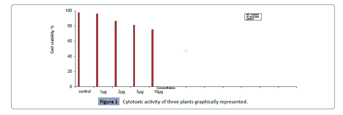
Cytotoxic activity of three plants extracts were carried out against A549 cell line at different concentrations to determine the IC50 (50% growth inhibition) by MTT assay. Results of different concentrations of plant extracts including (control, 1 μg/ml, 2 μg/ ml, 5 μg/ml, 10 μg/ml) are tabulated in Figure 1, and graphically represented in Figure 1.

Health-Science-journal-Cytotoxic

Figure 1: Cytotoxic activity of three plants graphically represented.

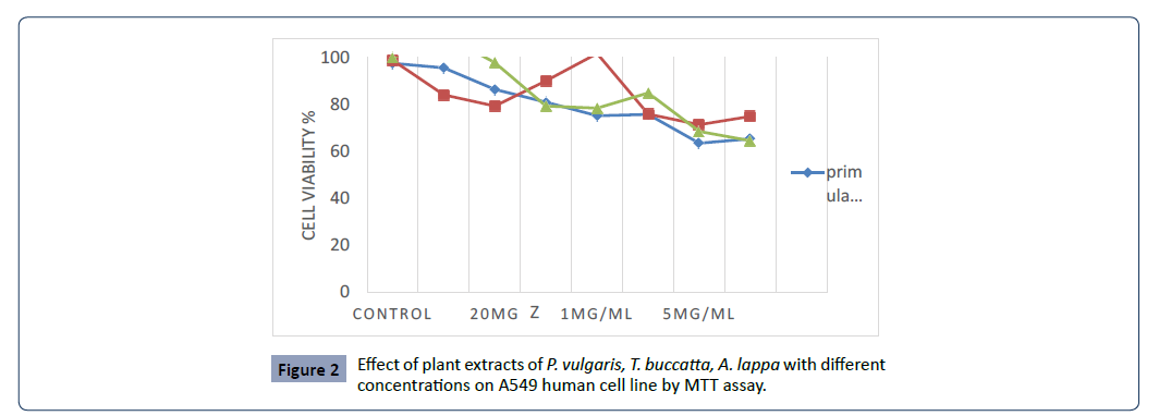
MTT assay of primula vulgaris L. shows significant effect on A549 cell in concentration range between 1 μg/ml to 5 μg/ml, compared with control. MTT assay of primula vulgaris L. shows significant effect on A549 cell in concentration range between 1 μg/ml to 5 μg/ml, compared with control. The highest cytotoxicity of this extract against A549 cell was found in 1 μg/ml and 1.5 μg/ ml concentrations with 51.45 and 56.49 percent of cell growth inhibition. It was found that the percentage of growth inhibition to be increasing with increasing concentration of test compounds and IC50 value of this assay was 1.01 μg/ml, however the IC50 of Taxus bucatta extract was about 0.1 μg/ml & the Paclitaxel with IC 50 0.01 μg/ml , a comparative analysis as shown in Figure 2.

Health-Science-journal-Effect

Figure 2:Effect of plant extracts of P. vulgaris, T. buccatta, A. lappa with different concentrations on A549 human cell line by MTT assay.

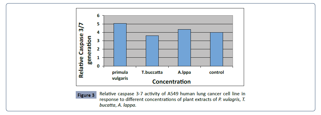
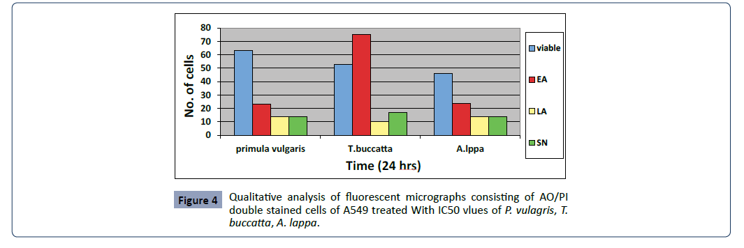
Cytotoxic activity of three plants extracts were carried out against A549 cell line with IC50 (50% growth inhibition) to determine the morphological, apoptotic changes, mRNA expression by the following methods- acridine orange (AO)/propodium iodide (PI) double staining fluorescence microscope, caspase 3-7 activity assay, real time RT-PCR as given in Figures 3 and 4.

Health-Science-journal-Relative

Figure 3:Relative caspase 3-7 activity of A549 human lung cancer cell line in response to different concentrations of plant extracts of P. vulagris, T. bucatta, A. lappa.

Health-Science-journal-Qualitative

Figure 4: Qualitative analysis of fluorescent micrographs consisting of AO/PI double stained cells of A549 treated With IC50 vlues of P. vulagris, T. buccatta, A. lappa.

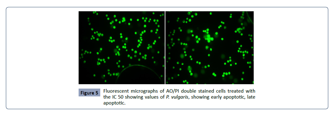
Fluorescent microscopy was conducted to study morphological changes of cell death mode induced by P. vulgaris, A. lappa, T. buccatta extracts, Paclitaxel after 12, 24, 48 and 72 h. Viable cells displayed green fluorescence with the appearance of circular cell; intact nucleus. The early apoptotic cells have fragmented DNA, which gives several green coloured nuclei and cell blebbing. Late apoptotic and necrotic cell’s DNA would be fragmented and stained orange red. No necrosis was obtained in P.Vulagris but the apoptotic effect was significantly observed however necrosis was attained by Taxus buccatta, paclitaxel. The percentage of apoptotic cells in untreated cells slightly increased from 0.06% after 24 h to 5.33% and 7% after 48 and 72 h, respectively. The percentage of apoptotic cells increased rapidly from 37% after 24 h to 53% and 63% after 48 h and 72 h respectively as given in Figure 5.

Health-Science-journal-Fluorescent

Figure 5: Fluorescent micrographs of AO/PI double stained cells treated with the IC 50 showing values of P. vulgaris, showing early apoptotic, late apoptotic.

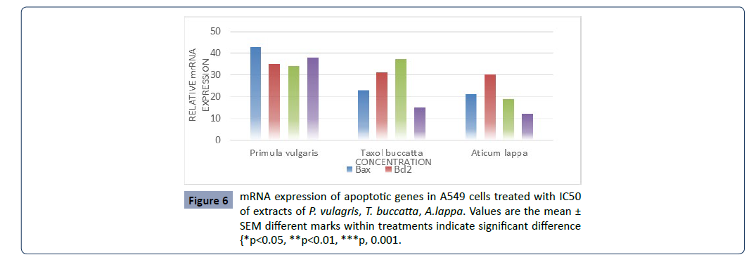
The expression of genes associated with apoptotic cell death, including the tumor suppressor p53, pro-apoptotic Bax, the antiapoptotic Bcl-2 and Fas gene in A549 cells was determined by RT-PCR. Results show that P. vulgaris, T. buccatta and A. lappa extracts, significantly increased the expression of p53, Bax, Fas compared to control. However the expression of Bcl-2 was not decreased compared to that of the control (Figure 6).

Health-Science-journal-mRNA

Figure 6:mRNA expression of apoptotic genes in A549 cells treated with IC50 of extracts of P. vulagris, T. buccatta, A.lappa. Values are the mean ± SEM different marks within treatments indicate significant difference {*p<0.05, **p<0.01, ***p, 0.001.

Taken together, the P. vulgaris extracts were more effective in inducing apoptosis through the regulation of p53, Bax and Fas expression.

Review of Literature

Taxanes currently known to suppress and inhibit cell growth, differentiation and proliferation in indefinitely known cancer cell Either it be in experimental or clinical trials, their mechanisms related to decrease cell growth has been thoroughly appreciated by everyone including patients in the oncological field [40]. Inspire of their excellent anti- tumor/anti-cancer activity an emergence of resistance to chemotherapeutic agents remains a major problem in the treatment of patients especially lung cancer, despite the fact that these patients usually have a good initial response to chemotherapy, as number of genetic alterations associate d with drug resistance in lung cancer are known, including the deregulated expression of proteins involved in drug transport and activity [17], and in cell cycle checkpoint control [18]. An alternate and more appropriate therapy needs to be carried out to delineate & prevent chemo resistance. In the present study, results indicate that docetaxel, paclitaxel and prunella vulgaris induce cytotoxicity in cancer cells in a timeand dose‐dependent manner, which is consistent with results of previous studies [33-36] In addition several studies [37, 38] Possible mechanisms suggested the inhibition of the c-Jun N-terminal kinase (JNK) pathway and the Akt pathway [39,40]. P.

vulgaris inhibits the proliferation of human cancer cell lines [41] including human oesophageal cancer cell line Eca 109 liver cancer cell line HepG2, cervical cancer Hela cell, and stomach cancer MKN 45 cell line Ethanol extract of P. vulgaris was found to inhibit colon cancer cell line HT-29 by arresting the cell cycle at the G1/S checkpoint and reducing the expression of pro-proliferative cyclin D1 and cyclin-dependent kinase 4 (CDK4) at the transcriptional and translational level P. vulgaris has also been shown to have combinatorial effects with other agents. P. vulgaris extracts enhanced the effects of paclitaxel (TAX) and adriamycin (ADM) on inhibiting cell growth of cancer cells A combination of P. vulgaris and Cremastra appendiculata exhibited an enhanced effects in inhibiting the growth of thyroid cancer cell line along with downregulation of the c-myc expression.

In addition to P. vulgaris extracts, some of its ingredients have also been examined for anti-cancer activities. Triterpenoicacids, a component of P. vulgaris exhibited strong cytotoxic activity against human lung cancer cell line A459 Triterpenoic acid isolated from P. vulgaris has been shown to inhibit cell growths of various human cancer cell lines, namely, A549 cell lines, SK-OV-3 (ovarian cancer cell), SK-MEL-2 (skin melanoma), and HCT 15 (colon cancer cell). Ursolic acid one of the most abundant triterpenoic acids in P. vulgaris, showed inhibitory effect on colon cancer cell lines HCT-15 and DLD-1. Ursolic acid also reduced proliferation in many other tumor cell lines, like human leukemic cell line HLmouse melanoma cell line B16 human breast MCF7 Oleanic acid, an active component of P. vulgaris inhibited the proliferation of HT-29 cells in dose-dependent manner through the mechanism of G0/G1 checkpoint arrest. Oleanolic acid also exhibited strong anti-proliferation activity against human lung SPC-A-1 cells.

Regulation of cell cycle progression and cell cycle arrest

Taxanes achieve favourable apoptotic outcomes by ability of binding to microtubules-stabilize them, inhibit depolymerisation, interfere with the G2/M phases (64) which are achieved by blocking the cell cycle during mitosis in the transition from prometaphase to metaphase and hence, induce apoptosisprogrammed cell death confirmed through cytometry studies which is a crucial checkpoint in cancer treatment. Moreover, they also initiate a whole cascade of cell death pathways P. vulgaris has also been shown to induce cell cycle arrest at various checkpoints in cancer cells. After thyroid carcinoma cell line SW579 was treated with P. vulgaris, the proportion of cells in the S phase was observed to be reduced, while those in the G0/G1 phase was significantly increased when compared to the control group. In another study, the ethanol extract of P. vulgaris arrested cells at the G1/S checkpoint in human colon carcinoma cells and inhibited the expression of both cyclin D1 and CDK4. Rutin, one of the flavonoids from P. vulgaris, showed anti-tumor effect against human neuroblastoma LAN-5 cells by inducing G2/M cell cycle arrest and apoptosis. Ursolic acid, another component of P. vulgaris, was shown to block B16 mouse melanoma cell line in G1 phase which is in accordance with the present study. These reports suggest that P. vulgaris is also capable of inducing cell cycle arrest in various cancer cell lines.

Induction of apoptosis

Taxanes induce apoptosis by regulating c-Raf-1 kinase an important mediator of programmed cell death which is somehow concentration dependent; increase stabilization of protein by induction of wild-type p53 and p21WAF1 and down regulate the proto- oncogene c-myc thus, promoting apoptosis. Amongst the whole myriad of genes enhancing taxanes induced apoptosis, there is the Bcl-2 family where it is speculated that taxanes can interact and induce cytotoxicity via phosphorylation of Bcl-xl (B-cell lymphoma-extra-large) and Bcl-2(B-cell lymphoma 2)/BAX (Bcl-2–associated X protein) which are members of the apoptosis regulator proteins they are also known to cause resistance in tumor cells but, nevertheless, play a pivotal role in both breast and prostate cancer treated regimens. However, recently Bcl- 2 has been found to enhance taxanes chemo sensitivity in some solid tumors therefore, changing it from a protector to a killer which proves to be a completely novel strategy and a plus in cancer battle. P. vulgaris and its components have also been shown to induce apoptosis in a variety of cancer cell lines (including Raji cells, SGC- 7901, SW 579, Eca 109, EL-4, Jurket cells, PANC-1, T24, HepG2, HT29, A549, MKN-45, and Hela cells Several phytochemicals from P. vulgaris including oleanic acid ursolic acid rosmarinic acid and caffeic acid have also been shown to either induce or promote apoptosis in cancer cells. Mechanisms suggested by several studies are both the up-regulation of the expression of p53,Bax Fas Bad caspase 3 and caspase 9 as well as down-regulation of the expression of c-myc Bcl-2 Mcl and Bclxl Other mechanisms have been suggested are the inhibition of mitogen-activated protein kinase/extracellular signal-regulated kinase (MAPK/ERK) pathway the mitochondrial pathway the nuclear transcription factor NF-κB pathway, and the intracellular generation of reactive oxygen species (ROS) The present study highlighted that P. vulgaris var. lilacina affected the expression of genes associated with apoptotic cell death, including the tumor suppressor p53,pro-apoptotic Bax, the anti-apoptotic Bcl-2 and Fas genes in A549 cells.

P53-mediated apoptosis primarily occurs through the intrinsic apoptotic program It was reported that p53 induces apoptosis by either increasing transcriptional activity of proapoptotic genes such as Bax or suppressing the activity of the anti-apoptotic genes of the Bcl-2 family Our data show that P. vulgaris var. lilacina significantly increased the expression of p53, Bax and Fas compared to the control. However, the expression of Bcl-2 was not decreased compared to that of the control (Figure 6). Therefore, the treatments altered the expression of Bax/Bcl-2, resulting in a shift in their ratio favouring apoptosis. Several other groups have shown in various cancer cell lines that P. vulgaris var. lilacina can lead to cell death by inducing apoptosis through regulation of p53 and Bax/Bcl-2 expression In our study, the resulting elevation in p53 and Bax protein expression in lung cancer cells is consistent with our earlier proposed involvement of p53 and Bax-related response systems. Taken together, we suggest that P. vulgaris var. lilacina induce apoptosis through the regulation of p53, Bax, and Fas expression.

Conclusion

P. vulgaris has been extensively used in China both independently and as a part of a multi-modal approach to treat cancer patients with standard chemotherapy. P. vulgaris appears to target multiple signalling pathways and has a complex mechanism of action. The complexity of the herb may be a key element of its therapeutic or preventive effectiveness. However, the pleiotropic effects that it causes make determining definitive targets for future pharmaceutical development more challenging. Based on its strong efficacy in both pre-clinical model systems and in a number of clinical trials with limited toxicity or adverse effects, further studies should focus on characterizing P. vulgaris as a promising cancer chemo preventive agent.

References

  1. Mathers CD, Boschi-Pinto C (2001) Global burden of cancer in the year 2000: Version 1 estimates. Geneva, World Health Organization. GBD 2000 Draft Methods Paper.
  2. Indexed at, Google Scholar, Crossref

  3. Hong WK, Sporn MB (1997) Recent advances in chemoprevention of cancer. Science (Wash. DC), 278: 1073-1077.
  4. Indexed at, Google Scholar, Crossref

  5. Newman DJ, Cragg GM, Snader KM (2003) Natural products as sources of new drugs over the period 1981–2002. J Natural Prod 66: 1022-1037.
  6. Indexed at, Google Scholar, Crossref

  7. Mqoqi N, Kellett P, Sitas F, Jula M (2004) Incidence of histologically diagnosed cancer in South Africa, 1998-1999. National Cancer Registry South Africa, Johannesburg 1-96.
  8. Google Scholar

  9. Koul PA, Koul SK, Sheikh MA, Tasleem RA, Shah A (2010) Lung cancer in the Kashmir valley. Lung India 27: 131- 137.
  10. Indexed at, Google Scholar, Crossref

  11. Time trend in cancer incidence rates 1982-2005 (2009) National cancer registry programme, ICMR.
  12. Google Scholar

  13. Sharma RG, Kumar R, Jain S, Jhajhria S, Gupta N et al. (2009) Distribution of malignant neoplasms reported at different pathology centres and hospitals in Jaipur, Rajasthan. Indian J cancer 46: 323-330.
  14. Indexed at, Google Scholar, Crossref

  15. Malothu N, V eldandi U, Y ellu N, Devarakonda R, Y adala N (2010) Pharmacoepidemiology of oral cancer in Southern India. Internet J Epidemiol 8: 1.
  16. Indexed at, Google Scholar

  17. Tannock IF, de Wit R, Berry WR, Horti J, Pluzanska A et al. (2004) Docetaxel plus prednisone or mitox- antrone plus prednisone for advanced prostate cancer. N Engl J Med 351: 1502-1512.
  18. Indexed at, Google Scholar, Crossref

  19. Khan Y, Bobba R (2003) Cancer in India: An Overview. GOR 5: 93-96.
  20. Indexed at, Google Scholar

  21. Di Cosimo S, Ferretti G, Fazio N, Silvestris N, Carlini P et al. (2003) Docetaxel in advanced gastric cancer: review of the main clinical trials. Acta Oncologica 42: 693-700.
  22. Indexed at, Google Scholar, Crossref

  23. Roth AD, Ajani J (2003) Docetaxel-based chemotherapy in the treatment of gastric cancer. Ann Oncol 14: 41-44.
  24. Indexed at, Google Scholar, Crossref

  25. Nabell L, Spencer S (2003) Docetaxel with concurrent radiotherapy in head and neck cancer. Semin Oncol 30: 89-93.
  26. Indexed at, Google Scholar, Crossref

  27. Wani MC, Taylor HL, Wall ME et al (1971) Plant antitumor agents. VI. The isolation and structure of taxol, a novel antileukemic and antitumor agent from Taxus brevifolia. J AM Chem Soc 93: 2325-27.
  28. Indexed at, Google Scholar, Crossref

  29. Bissery MC, Guénard D, Guéritte-Voegelein F et al (1991) Experimental antitumor activity of taxotere (RP 56976, NSC 628503), a taxol analogue. Cancer Res 51: 4845-52.
  30. Indexed at, Google Scholar

  31. Patel N, Chatterjee SK, Vrbanac V et al (2010) Rescue of paclitaxel sensitivity by repression of Prohibitin1 in drug- resistant cancer cells. PNAS 107: 2503-2508.
  32. Indexed at, Google Scholar, Crossref

  33. Doyle LA (1993) Mechanisms of drug resistance in human lung cancer cells. Semin Oncol 20: 326-337.
  34. Indexed at

  35. Bodner SM, Minna JD, Jensen SM, D’Amico D, Carbone D et al. (1992) Expression of mutant p53 proteins in lung cancer correlates with the class of p53 gene mutation. Oncogene 7:743-9.
  36. Indexed at, Google Scholar

  37. Hennenfent KL, Govindan R (2006) Novel formulations of taxanes: a review. Old wine in a new bottle? Ann Oncol 17: 735-749.
  38. Indexed at, Google Scholar, Crossref

  39. Gudena V, Montero AJ, Glück S (2008) Gemcitabine and taxanes in metastatic breast cancer: a systematic review. Therapeutics and Clinical Risk Management 4: 1157-1164.
  40. Indexed at, Google Scholar, Crossref

  41. Ahmad I, Mehmood Z, Mohammad F (1998) Screening of some Indian medicinal plants for their antimicrobial properties. J Ethnopharmacol 62: 183-193.
  42. Indexed at, Google Scholar, Crossref

  43. Datta BK, Rahman I, Das TK (1998) Antifungal activity of Indian plant extracts Mycoses 41: 535-536.
  44. Google Scholar

  45. Abo KA, Adeyemi AA, Adeite DA (2000) Ethnobotanical survey of plants used in the treatment of infertility and sexually transmitted diseases in southwest Nigeria. Afr J Med Sci 29: 325-327.
  46. Indexed at, Google Scholar

  47. Graf J (2000) Herbal anti‐inflammatory agents for skin disease. Skin Therapy Lett 5: 3-5.
  48. Indexed at, Google Scholar

  49. Ankli A (2002) Yucatec Mayan medicinal plants: evaluation based on indigenous uses. J Ethnopharmacol 79: 43-52.
  50. Google Scholar, Crossref

  51. Neto C (2002) Antibacterial activity of some Peruvian medicinal plants from the Call Jon de Huaylas. J Ethnopharmacol 79: 133-138.
  52. Indexed at, Google Scholar, Crossref

  53. Ahmedullah M (1999) Red Data book of Indian Plants. Botanical Survey of India
  54. Commonly used herbal medicines in the United States: a review. Am J Med 116: 478-485.

    Google Scholar, Crossref

  55. Samant SS, Dhar U, Palni LMS (1998) Medicinal Plants of Indian Himalaya: Diversity Distribution Potential Values. Nainital: Gyanodaya Prakashan.
  56. Indexed at, Google Scholar

  57. Samant SS, Butola JS and Sharma A (2007) Assessment of diversity, distribution, conservation status and preparation of management plan for the medicinal plants in the catchment area of Parbati Hydro-Electric Project Stage III in North Western Himalaya. J Moun Sci 4: 34-56.
  58. Google Scholar

  59. Cardellina II JH, RW Fuller, WR Gamble C, Westergaard J Boswell (1999) Evolving strategies for the selection dereplication and prioritization of antitumor and HIV-inhibitory natural products extracts. In: Bioassay Methods in Natural Product Research and Development Bohlin L and JG Bruhn, (Eds.). Kluwer Acad. Pub Dordrecht 25-36.
  60. Indexed at, Google Scholar, Crossref

  61. Riccardi A, Servidei T, Tornesello A, Puggioni P, Mastrangelo S et al. (1995) Cytotoxicity of paclitaxel and Docetaxel in human neuroblastoma cell lines. Eur J Cancer 31A: 494‐499.
  62. Indexed at, Google Scholar, Crossref

  63. Rosetti M, Zoli W, Tesei A, Ulivi P, Fabbri F et al. (2007) Iressa strengthens the cytotoxic effect of docetaxel in NSCLC models that harbor specific molecular characteristics. J Cell Physiol 212: 710‐716.
  64. Indexed at, Google Scholar, Crossref

  65. Shurin GV, Tourkova IL, Kaneno R, Shurin MR (2009) chemotherapeutic agents in noncytotoxic concentrations increases antigen presentation by dendritic cells via an IL‐12‐dependent mechanism. J Immunol 183: 137‐44.
  66. Indexed at, Google Scholar, Crossref

  67. Kim YJ, Lee CJ, Lee U, Yoo YM (2005) Tamoxifen‐induced cell death and expression of neurotropic factors in cultured C6 glioma cells. J Neurooncol 71: 121‐151.
  68. Indexed at, Google Scholar, Crossref

  69. Kim JH, Kim YH, Song GY (2014) Ursolic acid and its natural derivative corosolic acid suppress the proliferation of APC-mutated colon cancer cells through promotion of beta-catenin degradation. Food Chem Toxicol Int J Published Br Ind Biol Res Assoc 67: 87-95.
  70. Indexed at, Google Scholar, Crossref

  71. Juan ME, Planas JM, Ruiz-Gutierrez V, Daniel H, Wenzel U (2008) Antipoliferative and apoptosis-inducing effects of maslinic and oleanolic acids, two pent acyclic triterpenes from olives on HT-29 colon cancer cells. Br J Nutr 100:36-43.
  72. Indexed at, Google Scholar, Crossref

  73. Liu XK, Wang L, Zhang MZ (2010) Involvement of JNK and caspase-3 in human lymphoma cell apoptosis induced by Prunella vulgaris. Zhonghua Yi Xue Za Zhi 90: 690-693.
  74. Indexed at, Google Scholar

  75. Liu XK, Wang L, Zhang MZ (2010) Roles of JNK pathway and inhibition of lymphoma cells by Prunella valgaris. Acad J Second Mil Med Univ 31: 452-457.
  76. Indexed at, Google Scholar

  77. Shi XQ, Lv XQ (2008) Mechanism of inhibition the human lymphoma cell growth by PV. China Pharm 12: 1038-1040.
  78. Indexed at, Google Scholar, Crossref

  79. Chen CY, Wu G, Zhang MZ (2008) Effect of Prunella vulgaris L extract on proliferation of human Burkitt lymphoma cell line Raji. J Zhengzhou Univ (Med Sci) 43: 1189-1192.
  80. Indexed at, Google Scholar

  81. Fu XR, Zhang MZ, Liu HM (2008) Effect of extract from Prunella Vulgaris on Jurket cell apoptosis. J Zhengzhou Univ (Med Sci) 43: 964-967.
  82. Google Scholar

Citation: Neelam D, Husain SA, Tabasum T, Maqbool lone M (2023) Efficacy of Taxol and Prunella vulgaris against Human Lung Cancer Cell Line-A549. Health Sci J. Vol. 17 No. 10: 1069.