Case Report - (2022) Volume 0, Issue 0
Fetus in Fetu
Luis A. Mejia-Vargas1*,
Juan Esteban Tafur Delgado2,
Sayne Sailet Gonzalez Alvarez3,
Villaveces Buelvas4,
Valentina Lobo Perez5,
Lucia Vellojin Olivares6,
Dayana Judith de la rosa Gonzalez7,
Carlos Mauricio Vergara lobo8 and
Julio Cesar Anichiarico Cecere9
1General Surgery, Clinica Santa Maria. University Teacher, Universidad de Sucre, Sincelejo, Colombia
2Third year Pediatric Resident, Universidad del Sinu, Colombia
3General Physician, Fundacion Maria Reina, Sincelejo, Colombia
4General Physician, Fundacion Maria Reina, Sincelejo, Colombia
5Intern Physician, Fundacion Santa Maria, Sincelejo, Colombia
6First Year Pediatric Resident, Universidad del Sinu, Colombia
7Epidemiologist, Universidad Autónoma de Bucaramanga, Colombia
8Pediatric Infectologist, Universidad de Buenos Aires, Argentina. University Teacher, Universidad de sucre, Colombia
9Pediatric Surgeon, Hospital San Jeronimo de Monteria, Colombia
*Correspondence:
Luis A. Mejia-Vargas, General Surgery, Clinica Santa Maria. University Teacher, Universidad de Sucre, Sincelejo,
Colombia,
Email:
Received: 30-May-2022, Manuscript No. Iphsj-22-12804;
Editor assigned: 01-Jun-2022, Pre QC No. Iphsj-22-12804 (PQ);
Reviewed: 15-Jun-2022, QC No. QC No. Iphsj-22-12804;
Revised: 20-Jun-2022, Manuscript No. Iphsj-22-12804(R);
Published:
27-Jun-2022, DOI: 10.36648/1791-809X.16.S7.955
Abstract
The fetus in fetus (FIF) is a rare congenital pathology with less than 200 cases reported in the literature worldwide, in which a pregnancy with monozygotic twins one of them develops normally, while the other atrophies and it is inside the developed twin. The most frequent location is retroperitoneal; however, multiple cases have been described in the sacrum, mediastinum, skull and neck. We present a case of a 17-day�old female patient diagnosed with a congenital abdominal mass. After the extraction, an anatomopathological analysis was performed. There were multiple limbs, vertebral column, anencephalic cranial cavity, and the diagnosis of FIF was concluded.
Keywords
Fetus in fetu, Pediatric surgery, Retroperitoneal, Teratoma
Introduction
Fetus in fetu is a rare congenital pathology, with an incidence of
1 in 500,000 births [1], in which there are 2 asymmetrical twins
and the parasitic twin develops abnormally within the body of the
host twin [2] This was discovered by Meckel in 1800 and defined
by Willis in 1953, as a mass containing vertebral axis, frequently
associated with other organs and limbs around this central axis
[3].
It is believed that the development of this is related to the
uneven division of totipotent cells in the blastocyst, this is how
two monozygous twins do not completely separate and are joined
by some area, which has been said to be the anastomosis of the
vitellin circulation. One of these twins grows healthy while the
other atrophies staying inside the twin healthy and completely
dependent on him. It is unknown why twins do not separate
properly [4, 6, 9], and this monozygous twin theory does not
explain all cases of FIF, including cases characterized by the
presence of multiple fetuses associated with a single host or an
FIF associated with a teratoma, however, this is supported by the
fact that studies of genetic markers and genotyping suggest that
host babies and their fetish masses are genetically identical [5].
The overall incidence is 1 in 500,000 live births. In most cases,
they are found in babies. Usually in FIF, the parasitic fetus is
anencephalic and cardiac, but the limbs are described in 82.5%
and the spine in 91% of cases [6]. The most common presentation
is abdominal, more specifically retroperitoneal (80%) along
the ventral midline, skull 8% and sacrococcygeal region 8% [7],
however, cases have been reported in mediastinum, intracranial,
sacrum, scrotum, neck and oral cavity [8], and tend to produce
mass effect.
The diagnosis is sometimes not easy, it can be done by means
of radiography, ultrasonography, tomography and magnetic
resonance which is gaining prominence due to its high resolution,
low irradiation and the non-use of a contrast medium [7, 9, 10],
in some cases they can be underdiagnosed, being reported as
teratoma, which is one of the differential diagnoses and whose
differentiation is given by the presence of a vertebral organization
with buds of the limbs and other organ systems [7], being of
great relevance due to the malignant potential of the latter, this
being refuted as a point of differentiation by some theories until
now without solid foundations.
Although FIF usually occurs during infancy and early childhood
before 18 months of age, a small number of case reports
of patients with FIF during childhood, early childhood, and
adulthood have been published [10].
Case Report
This is a 17-day-old female patient born because of cesarean
section with a prenatal diagnosis of intra-abdominal malformation
at the renal level and polyhydramnios, who at birth had a globose
abdomen. Abdominal ultrasound is performed that reports spaceoccupying
mass with probable cystic appearance at the level of
the left kidney, a scintigraphy is performed that shows kidneys
in a normal position, left kidney increased in size, with a welldefined
area of hypouptation that involves a large part of the kidney
in the upper and middle regions of it, with minimal uptake area in
the lower pole. She is taken to surgery to perform left nephrectomy,
with the following findings: cystic mass, retroperitoneal location,
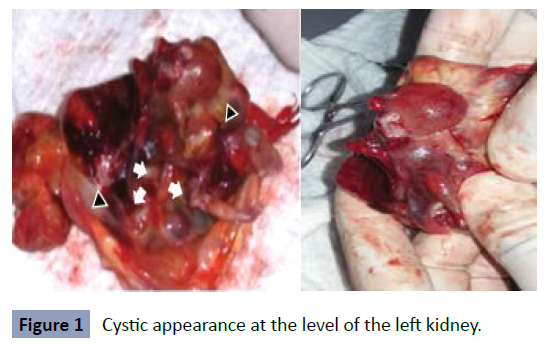
with content of fetoid structures as seen in (Figure 1). There is a left
kidney displaced by this mass towards the left iliac fossa, in addition
there was the presence of two vessels in the fetus. The two vessels
and the left kidney are preserved, mass is extracted finding in its
interior cetrino liquid as seen in image 2, in addition the presence
of a fetus with upper and lower extremities and two more joined
fetuses is evidenced forming a mass effect where only limbs are
distinguished, they are extracted and the sample is sent to
pathological anatomy study with report of fetus in fetu.
Figure 1: Cystic appearance at the level of the left kidney.
Discussion
Fetus in fetu is defined as the condition in which a fetus of
monozygous twins is incorporated during the separation process
into the body of the other twin. It is a pathology of low incidence with few articles worldwide; In Colombia there are few reported
cases, it is not clear if it is due to the low incidence of these or to
an underdiagnoses of it [3].
It is pathology with a complex diagnosis, which also requires a
high level of clinical suspicion. Among the differential diagnoses
we have mainly the teratoma, of which it has been suggested
that there is considerable overlap between the two conditions,
and that both could represent two aspects of the same entity at
various stages of maturation [11]. Mature teratoma may contain
well-developed structures and sufficiently resemble a fetus,
sometimes also called a fetiform teratoma.
The main characteristics to make a differentiation between
both diagnoses are in the presence of a spine as a central
axis, development of limbs, presence of anencephalic cranial
cavities and in some cases organification. There are other
theories that suggest that once the metameric cranio caudal
pattern is established, many structures can survive beyond the
disappearance of the rudimentary axis, and thus accommodating
the concept of FIF as an organized form of teratoma, relevant in
the prognostic value given the risk of malignancy of the latter
[12]. However, as of 2000 about 90 cases of FIF had been reported
of which 91% of cases had a spine, 82.5% had limbs, 55.8% had
a central nervous system, 45% had a gastrointestinal tract and
40% had vessels. In all cases, the fetus was anencephalic and the
lower limbs were more developed than the upper limbs. Thus,
82.5% of reported FIF cases fit one or both definitions offered by
Willis and Gonzalez-Crussi. (2001) [13].
The FIF presents mostly retroperitoneal localization and the
diagnosis is made during fetal development, birth and childhood,
on the other hand, the fetiform teratoma is located in the ovaries
of women of childbearing age in most cases [14].
In this case, a 17-day-old patient with a prenatal diagnosis of
polyhydramnios and mass in the left kidney was presented. With
the help of imaging studies, the diagnosis of cystic-looking mass
with solid-liquid content inside is carried out, after its extraction
multiple limbs are identified, with cartilaginous cavities that
denote the shell, without brain content. In the pathological study
of the sample in fresh and after fixation with formaldehyde,
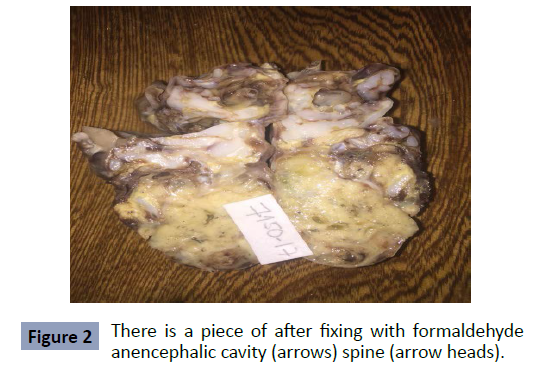
where the presence of a central axis is described as seen in image
3. Analyzing these characteristics and the criteria described
by Willis in 1953, the diagnosis of FIF is made, ruling out the
differential of fetish teratoma (Figure 2).
Figure 2: There is a piece of after fixing with formaldehyde
anencephalic cavity (arrows) spine (arrow heads).
REFERENCES
- Suhas A, Bibekanand J, Mrudula K (2017) Fetus in Fetu Case Report and Brief Review of Literature on Embryologic Origin, Clinical Presentation, Imaging and Differential Diagnosis. Pol J Radiol 82:46-49
Indexed at, Google Scholar, Crossref
- Virginia G, Rosales C, Tome E, Godoy C, Rodriguez H (2021) Fetus In Fetu: case clinic estudiado con tomographic tridimensional. Rev Med Hondur 89:S1-68.
Google Scholar
- Carmona Zenen Siosi A, Cabeza M (2016) Fetus in fetu: reporte de caso Horiz Med 16:63-67.
Indexed at, Crossref
- Lugones M, Ramirez Ma (2013) Rev Cubana Obstet Gynecol 39:63-68.
Google Scholar
- Ji Y, Song B, Chen S, Jiang X, Yang G (2015) Fetus in Fetu in the Scrotal Sac: Case Report and Literature Review. J Med 94:e1322.
Indexed at, Google Scholar, Crossref
- Marynczak L, Adamek D, Drabik G, Kwiatkowski S, Herman-Sucharska I (2014) Fetus in fetu: Medical curiosity-considerations based upon an intracranial located case. Childs Nerv Syst 30:357-360.
Indexed at, Crossref
- Ji Y, Chen S, Zhong L, Jiang X, Jin Set al. (2014) Fetus in fetu two case reports and literature. BMC Pediatrics 14:88.
Indexed at, Google Scholar
- Huang Y, Pan H (2017) Case Report A fetus with a mass in the oral cavity: a rare case of oral immature teratoma. Int J Clin Exp Pathol 10:7890-7892.
Indexed at, Google Scholar
- Yaacob R, Zainal Mokhtar A, Abang Jamari D, Jaafar N (2017) the entrapped twin: a case of fetus-in-fetu. BMJ Case Reports.
Indexed at, Google Scholar, Crossref
- Mustafa G Mirza B, Iqbal S Sheikh A (2012) A case of fetus in fetu. APSP J Case Rep 3:9.
Indexed at, Google Scholar
- Santos H, Furtado A, Tavares H, Joao A, Ferraz L et al. (2008) Caso Clinic Masa prenatal retroperitoneal teratoma o fetus in fetu. Cir Pediatr 21:185-187.
Indexed at, Google Scholar
- Spencer R (2001) Parasitic Conjoined Twins: External, Internal (Fetuses in Fetu and Teratomas), and Detached. Clinical Anatomy 14:428-444.
Indexed at, Google Scholar, Crossref
- Mills P, Bornick W, Morales J, Allen E, Gilbert-barness P (2001) Ultrasound prenatal diagnosis of fetus in fetu. Ultrasound obstet Gynecol 18:69-71.
Indexed at, Google Scholar, Crossref
- Traisrisilp K Srisupundit K, Suwansirikul S (2010) intracranial fetus-in-fetu with numerous fully developed organs.
Google Scholar, Crossref
Citation: MejIa-Vargas LA, Delgado JET,
Álvarez SSG, Buelvas MAV, Pérez VL, et al.
(2022) Fetus in Fetu. Health Sci J. Vol. 16 No.
S7: 955.


