Introduction
|
| Important themes are met in the long literature of bodily cancer colonization. It included a unilaterally disposed case dating to 1879 [1]. After the passage of a century in 1975, Ariel’s own encounter [2] was in someone in whom there were “innumerable” pigmented deposits and yet “The left side of the body remained completely inviolate [2].” “It was,” he had declared, “as if an imaginary line had been drawn vertically front and back to repel any invasion from right to left.” Personal interest in such dimensional data led to my final year publication based on the topographical analysis of 1000 cases of lung cancer. It appeared in the British Journal of Cancer in 1957 [3]. Thereafter, in the 1958 Edition of the famous Muir’s Text-Book of Pathology, Prof Cappell remarked thus: “Onuigbo, working in my Department, has shown that spread to the adrenals is predominantly to the ipsilateral side and this distribution indicates the lymphatics rather than the arterial blood stream as the usual route of metastases [4].” Next,instructive autopsy data were added from other Medical Schools during safaris undertaken in Scotland as follows: |
|
Examples of unilateral spread of lung cancer in Scotland
|
| Three cases are illustrative. They are illumined with italics for both the primary origins and the various colonies as follows: |
| i Glasgow Royal Infirmary (PM 8/1945). Left lung origin. On the under aspect of the left cerebellar lobe there is a secondary tumour nodule. There is also a single secondary tumour nodule in the left lobe of the liver and another in the left adrenal. |
| ii Edinburgh Royal Infirmary (PM 6/1946). Left lung origin. The lymph nodes in relation to the left hilum are replaced by cancerous tissue. Situated in the lower anterior aspect of the left lobe of the liver is a solitary spheroidal tumour deposit. The left adrenal contains tumour tissue. In the brain, a very large mass of tumour is situated in the left frontal and pariental lobes |
| iii Aberdeen Royal Infirmary (PM 92/1952). Right lung origin. Two small tumour nodules are seen, on section, in the right frontal lobe; a similar nodule is found in the right occipital lobe. On section, numerous secondary deposits of varying size are found scattered throughout the liver, more especially in the right lobe. The right adrenal is extensively invaded and weighs 130 gm. The left is free. |
Related Patterns
|
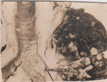
| Undoubtedly, these pointed patterns are, as Professor Scarf [5] concluded, a reflection of the grafting experiments done by Nature in man. In this context, the prevailing panorama was delineated personally with Scottish necropsy series which actually continued for years. The latest was entitled “The prevalence of endobronchial metastases in lung cancer in Scotland [6].” Thus, Figure 1 depicts whitish left lung tumor sheathing the lower trachea and showing coalesced endobronchial metastases with discrete extension just across the carina. This was a sequel to my possession of the copies of the 100 autopsies performed personally during my Postgraduate Training Program at the University Department of Pathology based at the Glasgow Western Infirmary. Incidentally, that Institution was aptly described historically in the Bulletin of the History of Medicine [7] as being second to none in Britain. |
| The postmortem materials were obtained with the mono-block formalin-fixation method whose enduring feature has been the long preservation of tissues from the neck to the pelvis [8]. Incidentally, although there is now the predatory problem of who takes possession of laboratory specimens [9], those packaged preciously by my Master were shipped by sea to me at Enugu, my base in the Capital City of the Eastern Region of Nigeria which is deep in the Dark Continent! |
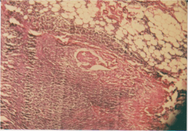
| In this context, the clear colonization patterns repeatedly pointed to the need to trace the footsteps of lymph-borne cancer cells all the way from the lung to, say, the abdominal lymph nodes [10]. In particular, I noted that the earliest deposit was present beneath the node’s convex border, i.e, where lymph flows in, (Figure 2). Accordingly, I theorized six times that new lymphatic vessels were being formed. Nowadays, this is being called “Lymphangiogenesis,” a phenomenon on which I have provided a review [11]. |
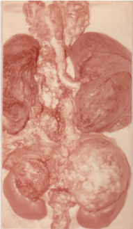
| I have also documented how contralateral neck nodes are involved [12], this being very important in scalene node surgery [13]. Furthermore, despite the long doubt spanning many decades [14,15], as to how cancer spreads to the pancreas and its abutting lymph nodes, my special mono-block maneuver confirmed the reality of the impact of lymphangiogenesis there [16]. For examples, Figure 3 draws attention to the definitely organized meanderings due to lymphangiogenesis. Thus, note how the right lung tumor metastasized upwards to the left sided neck nodes and downwards to the left sided adrenal gland, whereas the mediastinal node invasion continued to remain ipsilateral. |
| Among the laws of lymphatic metastasis, which I proposed in 1964, the 5th was on the “skip” phenomenon [17]. Nowadays, researches on it have continued. Thus, according to Kim [18], there are drainage patterns “such as skip metastasis and micrometastasis, (which) still remain the subject of investigation.” Likewise, Misthos [19] and associates analyzed its clinical significance and prognosis. What of its pathological significance? |
Discussion
|
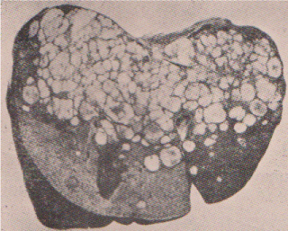
| The element of skipping presupposes choice of a particular soil. Actually, for centuries, the old masters questioned the attributes of “Site” and “Soil [20]. Recently, Kaplan and her group [21] wondered about this phenomenon in their research paper entitled “Preparing the ‘soil’: The premetastatic niche.” As they put it, “Current focus on cancer metastasis has centered on the intrinsic factors regulating the cell autonomous homing of the tumor cells to the metastatic site.” I am persuaded that this hitherto unknown “homing element” is the present proposed “Lymphogenous metastasis selectivity factor” (LMSF). For example, Figure 4. Illustrates the multiple deposits formed in a lung cancer case by way of the LMSF in the subjacent liver [22]. Surely, this pattern is not that of the haphazard distribution expected from the arterial system. |
Conclusion
|
| LMSF naturally directs positional activities during metastases. Consequently, what remains to be done is clear. It is, indeed, to hypothesize that this Factor is what is responsible for the welldirected natural phenomena which are traceable in lymphatic metastasis. In all probability, target therapy will result from recondite researches carried out on it, especially in the recently sprouting Translational Institutions [23]. |
Figures at a glance
|
 |
 |
 |
 |
| Figure 1 |
Figure 2 |
Figure 3 |
Figure 4 |
|
| |
| |

